Munir Sohbia, Khan Sohail Ahmed, Hanif Hina, Khan Maria
Sohbia Munir, Resident, Dow Institute of Radiology, Dow University of Health Sciences, Karachi, Pakistan.
Sohail Ahmed Khan, Assistant Professor, Dow Institute of Radiology, Dow University of Health Sciences, Karachi, Pakistan.
Pak J Med Sci. 2021 Jan-Feb;37(1):125-130. doi: 10.12669/pjms.37.1.2489.
To evaluate the diagnostic accuracy of magnetic resonance imaging (MRI) in detection of intra-axial gliomas in suspected cases keeping histopathology as gold standard.
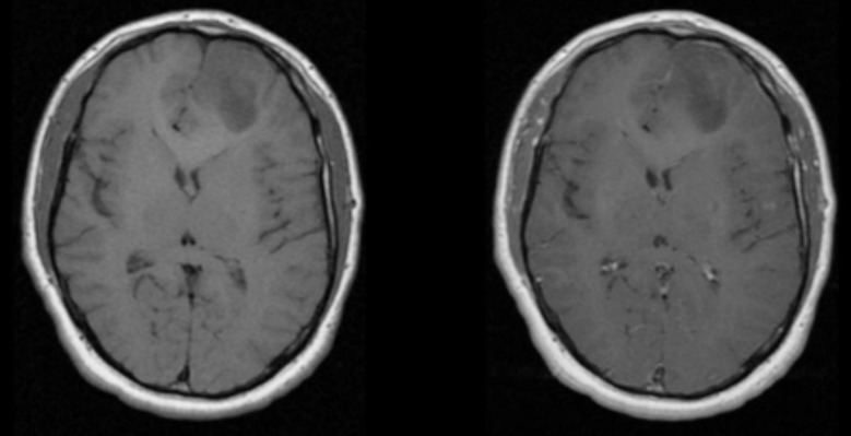
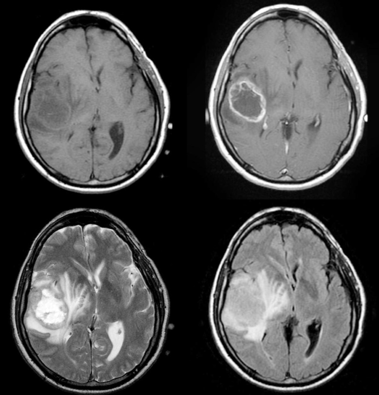
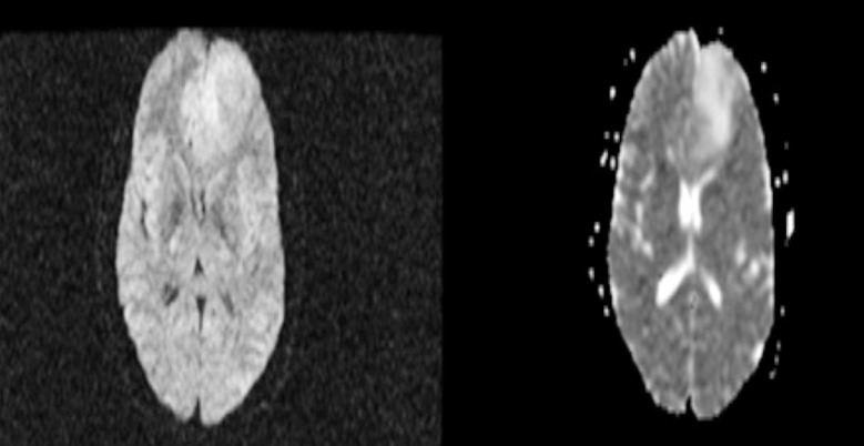
This cross-sectional study was conducted at Dow Institute of Radiology, DUHS from October 2017 - April 2018. Patients of either gender aged 30-70 years presenting with headache were included. Patients already diagnosed and referred for follow up were excluded. MRI was performed on 1.5T scanner by a trained MRI technician. T1, T2, FLAIR, diffusion weighted and T1 post contrast images were acquired and reviewed by two radiologists having more than five years post fellowship experience. Sensitivity, specificity, PPV, NPV and diagnostic accuracy of MRI for intraaxial gliomas was calculated taking histopathology findings as gold standard.
Mean age of the patient`s was 51.71 ±10.85 years. Positive intraaxial gliomas on MRI were observed in 123 (79.90%) patients while on histopathology, positive intraaxial gliomas were observed in 131 (85.10%) patients. Diagnostic accuracy of MRI in detection of intra-axial gliomas taking histopathology findings as gold standard showed sensitivity, specificity, positive predicted value (PPV), negative predicted value (NPV) and overall diagnostic accuracy as 89.31%, 73.91%, 95.12%, 54.84% and 87.01%.
MRI has high sensitivity, moderate specificity and high diagnostic accuracy in detection of intraaxial gliomas.
以组织病理学为金标准,评估磁共振成像(MRI)对疑似病例中轴内胶质瘤的诊断准确性。
本横断面研究于2017年10月至2018年4月在DUHS的陶氏放射学研究所进行。纳入年龄在30 - 70岁、出现头痛症状的男女患者。已确诊并转诊进行随访的患者被排除。由一名训练有素的MRI技术人员在1.5T扫描仪上进行MRI检查。采集T1、T2、FLAIR、扩散加权和T1增强后图像,并由两名具有五年以上 fellowship 后经验的放射科医生进行评估。以组织病理学结果为金标准,计算MRI对轴内胶质瘤的敏感性、特异性、阳性预测值(PPV)、阴性预测值(NPV)和诊断准确性。
患者的平均年龄为51.71±10.85岁。MRI显示123例(79.90%)患者存在阳性轴内胶质瘤,而组织病理学显示131例(85.10%)患者存在阳性轴内胶质瘤。以组织病理学结果为金标准,MRI检测轴内胶质瘤的诊断准确性显示敏感性、特异性、阳性预测值(PPV)、阴性预测值(NPV)和总体诊断准确性分别为89.31%、73.91%、95.12%、54.84%和87.01%。
MRI在检测轴内胶质瘤方面具有高敏感性、中等特异性和高诊断准确性。