Gong Wuyi, Yue Zhihang, Chu Haojun, Mi Xiaohui, Li Yongming
Shanghai Engineering Research Center of Tooth Restoration and Regeneration & Tongji Research Institute of Stomatology & Department of Orthodontics, Shanghai Tongji Stomatological Hospital and Dental School, Tongji University, No.399 Middle Yanchang Road, Jing'an District, Shanghai, 200072, China.
Stem Cell Res Ther. 2025 Jul 1;16(1):331. doi: 10.1186/s13287-025-04439-7.
Tensile force is a key regulator for condylar cartilage remodeling in the temporomandibular joint (TMJ), and this biomechanical characteristic underlies the mechanisms of mandibular growth modification in orthodontic practice. Cartilage stem/progenitor cells (CSPCs) in the superficial layer of condylar cartilage play an essential part in the development and remodeling of condylar cartilage. However, the regulatory role of tensile force on condylar CSPCs remains unclear. This study aimed to investigate the impact of tensile loading on condylar CSPCs and explore the molecular mechanisms within.
The mandibular advancement (MA) model was constructed to apply tensile force on the condylar cartilage in vivo. Flow cytometry and transcriptome sequencing were utilized to assess the percentage of CSPCs and gene expression in the superficial layer of rat condylar cartilage. Lineage tracing with cathepsin K (Ctsk) in mice was employed to trace the differentiation of CSPCs. 10% equibiaxial dynamic strain was loaded on rat CSPCs for cell stretching in vitro. GsMTx4 was used to inhibit the Piezo1 channel, and the calcium chelating agent BAPTA was used to block the Ca influx of rat CSPCs. siRNA was applied to knock down the protein kinase C alpha (Prkca) of rat CSPCs in vitro and in vivo.
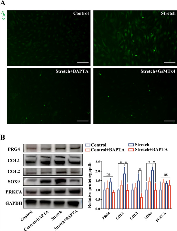
Cartilage thickening and a transient reduction of the CSPCs proportion in the superficial layer of the condylar cartilage were observed after 1 week of MA. The ratio of Ctsk and type II collagen double-positive cells climbed in the first week after MA, and 2 weeks later, the ratio of Ctsk and EdU double-positive cells rose. The expression level of chondrogenic-related genes, Piezo1, and Prkca was elevated in CSPCs after tensile loading. GsMTx4 and BAPTA could block the Ca influx into CSPCs caused by tensile stress. Furthermore, BAPTA and siPrkca could inhibit the stretch-induced chondrogenesis of CSPCs.
We uncovered that tensile stress could cause a transient shrinkage of the CSPCs pool in condylar cartilage, resulting from the accelerated chondrogenesis of CSPCs. Tensile force could promote the chondrogenic ability of CSPCs via the Piezo1-Ca-Prkca pathway. This study suggested a new regulatory route for mandibular growth modification in orthodontic practice.
张力是颞下颌关节(TMJ)髁突软骨重塑的关键调节因子,这种生物力学特性是正畸治疗中下颌生长改变机制的基础。髁突软骨表层的软骨干细胞/祖细胞(CSPCs)在髁突软骨的发育和重塑中起着重要作用。然而,张力对髁突CSPCs的调节作用仍不清楚。本研究旨在探讨拉伸加载对髁突CSPCs的影响,并探索其内在分子机制。
构建下颌前伸(MA)模型,在体内对髁突软骨施加张力。采用流式细胞术和转录组测序评估大鼠髁突软骨表层CSPCs的百分比和基因表达。利用组织蛋白酶K(Ctsk)在小鼠中进行谱系追踪,以追踪CSPCs的分化。对大鼠CSPCs施加10%的等双轴动态应变进行体外细胞拉伸。使用GsMTx4抑制Piezo1通道,并使用钙螯合剂BAPTA阻断大鼠CSPCs的钙内流。应用小干扰RNA(siRNA)在体外和体内敲低大鼠CSPCs的蛋白激酶Cα(Prkca)。
MA术后1周,观察到髁突软骨表层增厚,CSPCs比例短暂降低。MA术后第1周,Ctsk和II型胶原双阳性细胞的比例升高,2周后,Ctsk和EdU双阳性细胞的比例升高。拉伸加载后,CSPCs中软骨生成相关基因、Piezo1和Prkca的表达水平升高。GsMTx4和BAPTA可阻断拉伸应力引起的钙内流进入CSPCs。此外,BAPTA和siPrkca可抑制CSPCs的拉伸诱导软骨生成。
我们发现拉伸应力可导致髁突软骨中CSPCs池的短暂收缩,这是由于CSPCs软骨生成加速所致。张力可通过Piezo1-Ca-Prkca途径促进CSPCs的软骨生成能力。本研究为正畸治疗中下颌生长改变提出了一条新的调节途径。