Huang Jialiang, Yao Yongkang, Zhang Liao, Yan Chenxu, Guo Zhiqian
State Key Laboratory of Bioreactor Engineering, Key Laboratory for Advanced Materials and Joint International Research Laboratory of Precision Chemistry and Molecular Engineering Feringa Nobel Prize Scientist Joint Research Center Institute of Fine Chemicals Frontiers Science Center for Materiobiology and Dynamic Chemistry School of Chemistry and Molecular Engineering East China University of Science and Technology Shanghai China.
Smart Mol. 2024 Jul 4;2(3):e20240020. doi: 10.1002/smo.20240020. eCollection 2024 Sep.
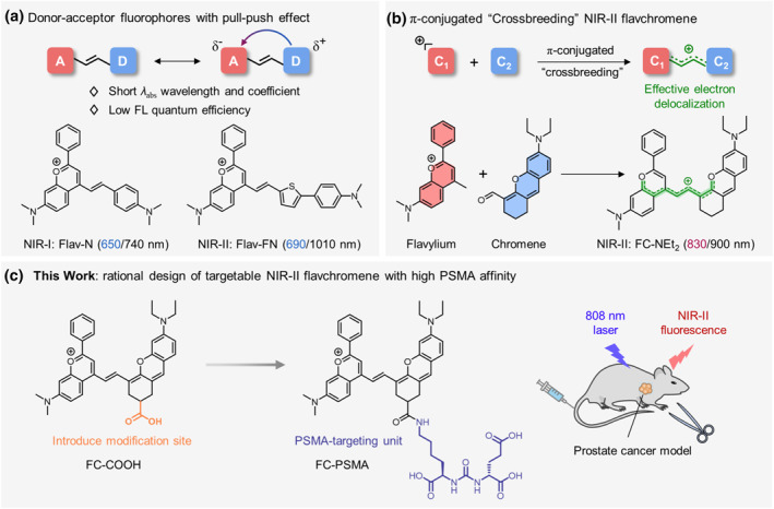
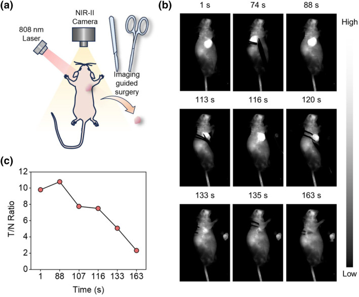
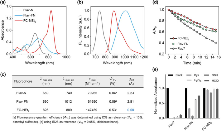
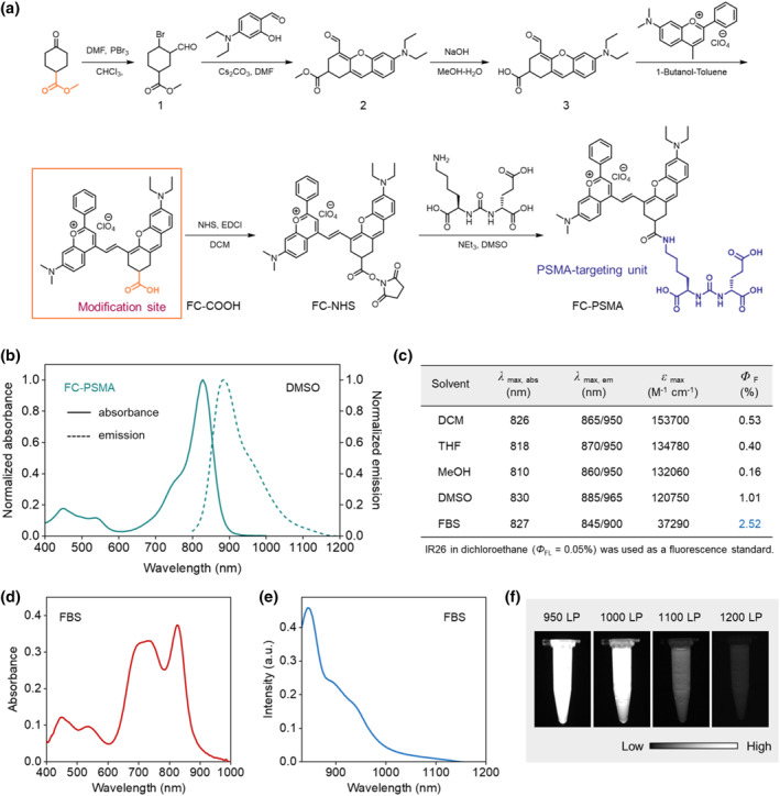
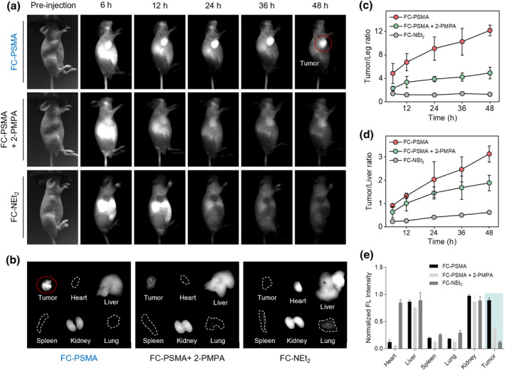
Prostate-specific membrane antigen (PSMA) is known to be overexpressed in prostate cancer (PCa). The development of precise and rapid imaging technologies to monitor PSMA is crucial for early diagnosis and therapy. Fluorescence imaging in the second near-infrared window (NIR-II) has emerged as a powerful tool for real-time tracking and in vivo visualization, offering high sensitivity and resolution. However, there is a lack of stable, bright and easy-to-implement NIR-II fluorescent probes for PSMA targeting. Herein, we presented a PSMA-targeting NIR-II fluorescent probe FC-PSMA based on π-conjugated crossbreeding dyed strategy that affords high stability, large extinction coefficient, and good brightness. As demonstrated, FC-PSMA displayed a high fluorescence quantum yield in fetal bovine serum (FBS). Following intravenous injection of FC-PSMA, the tumor-to-normal ratio of fluorescence intensity steadily increased over time, reaching a peak at 48 h (tumor-to-leg ratio = 12.16 ± 0.90). This advancement enables precise identification of PC through NIR-II fluorescence imaging, facilitating high-performance guidance for prostate cancer resection surgery.
已知前列腺特异性膜抗原(PSMA)在前列腺癌(PCa)中过表达。开发精确快速的成像技术来监测PSMA对于早期诊断和治疗至关重要。第二近红外窗口(NIR-II)荧光成像已成为实时追踪和体内可视化的有力工具,具有高灵敏度和分辨率。然而,缺乏用于靶向PSMA的稳定、明亮且易于实现的NIR-II荧光探针。在此,我们基于π共轭杂交染色策略提出了一种靶向PSMA的NIR-II荧光探针FC-PSMA,其具有高稳定性、大消光系数和良好的亮度。结果表明,FC-PSMA在胎牛血清(FBS)中显示出高荧光量子产率。静脉注射FC-PSMA后,荧光强度的肿瘤与正常组织比值随时间稳步增加,在48小时达到峰值(肿瘤与腿部比值 = 12.16 ± 0.90)。这一进展使得通过NIR-II荧光成像能够精确识别PCa,为前列腺癌切除手术提供高性能指导。