Hussein Sara M, Emanuels Andrew F, Leonel Luciano César P C, Lachman Nirusha, Peris Celda Maria, Morris Jonathan M, Sheahan Eric M, Martinez Erick O, Hanson Christian R, Sharaf Basel A
From the Division of Plastic and Reconstructive Surgery, Department of Surgery, Mayo Clinic, Rochester, MN.
Neural Engineering and Precision Surgery Laboratory, Mayo Clinic, Rochester, MN.
Plast Reconstr Surg Glob Open. 2025 Jul 9;13(7):e6972. doi: 10.1097/GOX.0000000000006972. eCollection 2025 Jul.
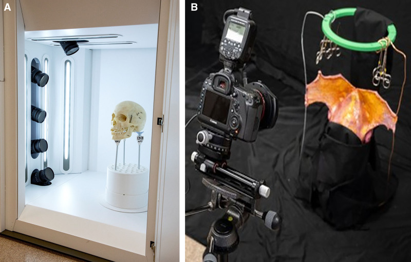
Anatomy is a critical component of surgical training; however, traditional resources-such as , , and anatomy applications-often fall short in delivering surgically relevant, 3-dimensional (3D) perspectives of facial anatomy. Although cadaveric dissection remains a valuable teaching tool, its accessibility is limited by cost, ethical concerns, and the lack of structured curricula, making it less feasible for ongoing surgical education. To address these limitations, this article introduced a novel educational approach that integrates 3D photogrammetry and virtual reality technology into the surgical anatomy curriculum for craniofacial and aesthetic surgery. Six fresh cadaveric specimens were meticulously dissected by a plastic surgeon to illustrate the intricate layered anatomy of the face and neck. At each stage of the dissection, a photogrammetry technique was used to capture 360-degree images using 5 digital single-lens reflex cameras alongside a 3D handheld camera. This approach not only ensured high-quality imaging but also facilitated the creation of detailed, lifelike virtual 3D models, enhancing the understanding of facial and neck anatomy within a surgical context. Additionally, we present some technical refinements of the 3D volume renderings to capture precise anatomical details during our dissections. Preliminary results from the use of this technology in anatomy courses and workshops indicated trainee engagement, a clearer grasp of the spatial anatomical relationships, and greater confidence in applying this knowledge to hands-on surgical procedures.
解剖学是外科培训的关键组成部分;然而,传统资源,如教科书、图谱和解剖学应用程序,在提供与手术相关的面部解剖学三维(3D)视角方面往往存在不足。尽管尸体解剖仍然是一种有价值的教学工具,但其可及性受到成本、伦理问题和缺乏结构化课程的限制,使得其在持续的外科教育中不太可行。为了解决这些局限性,本文介绍了一种新颖的教育方法,即将3D摄影测量和虚拟现实技术整合到颅面和美容手术的外科解剖学课程中。一位整形外科医生对六个新鲜尸体标本进行了细致的解剖,以展示面部和颈部复杂的分层解剖结构。在解剖的每个阶段,使用摄影测量技术,通过5台数码单反相机和一台3D手持相机拍摄360度图像。这种方法不仅确保了高质量的成像,还便于创建详细、逼真的虚拟3D模型,增强了在手术背景下对面部和颈部解剖结构的理解。此外,我们还介绍了一些3D体积渲染的技术改进,以便在解剖过程中捕捉精确的解剖细节。在解剖学课程和研讨班中使用该技术的初步结果表明学员参与度高、对空间解剖关系有更清晰的理解,并且在将这些知识应用于实际手术操作时更有信心。