Petriceks Aldis H, Peterson Ashley S, Angeles Miguel, Brown W Paul, Srivastava Sakti
Division of Clinical Anatomy, Stanford University School of Medicine, Stanford, CA, USA.
School of Health Sciences, Quinnipiac University, Hamden, CT, USA.
J Med Educ Curric Dev. 2018 Sep 17;5:2382120518799356. doi: 10.1177/2382120518799356. eCollection 2018 Jan-Dec.
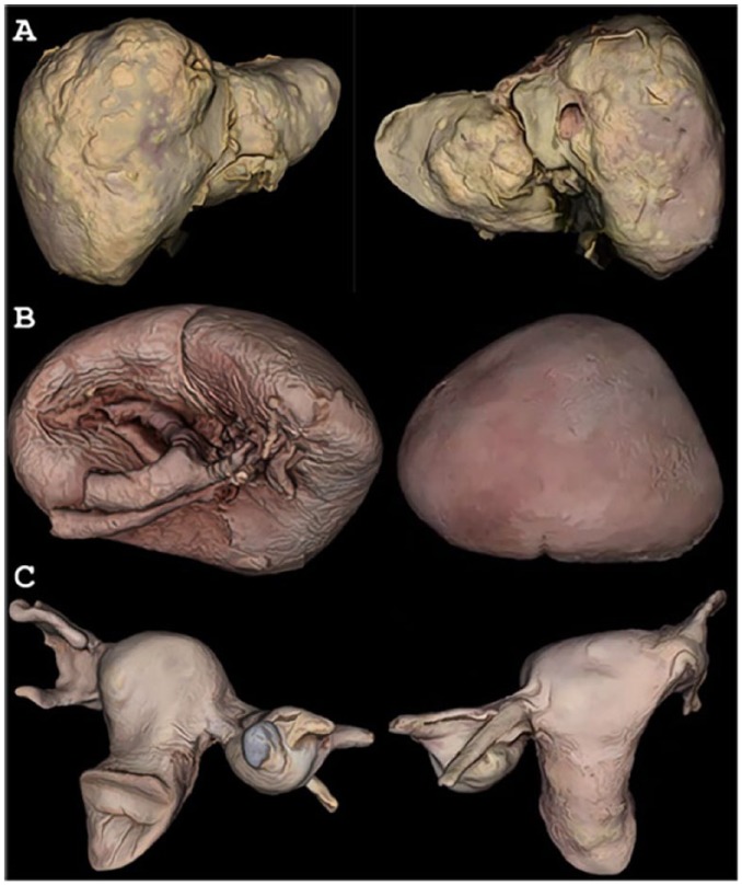
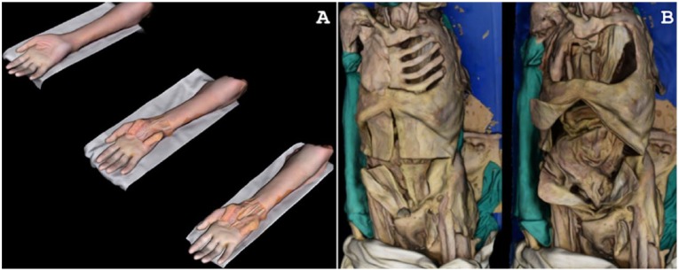
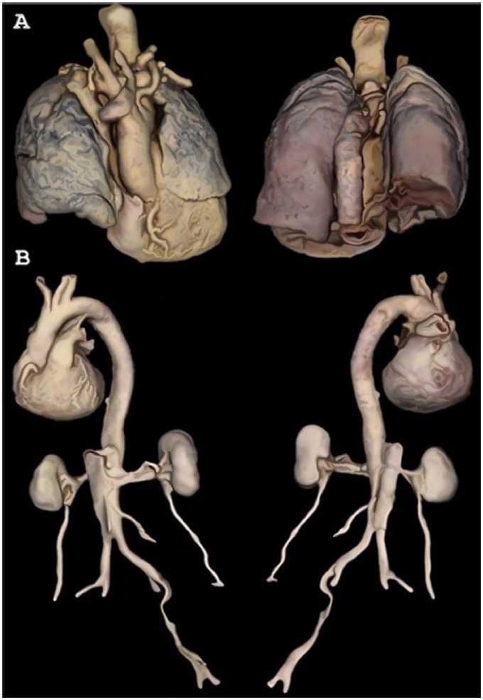
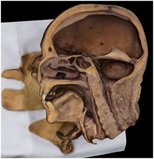
Cadaver-based anatomical education is supplemented by a wide range of pedagogical tools-from artistic diagrams, to photographs and videos, to 3-dimensional (3D) models. However, many of these supplements either simplify the true anatomy or are limited in their use and distribution. Photogrammetry, which overlaps 2-dimensional (2D) photographs to create digital 3D models, addresses such shortcomings by creating interactive, authentic digital models of cadaveric specimens. In this exploratory pilot study, we used a photogrammetric setup and rendering software developed by an outside group to produce digital 3D models of 8 dissected specimens of regional anatomy. The photogrammetrically produced anatomical models authentically and precisely represented their original specimens. These interactive models were deemed accurate and teachable by faculty at the Stanford University Division of Clinical Anatomy. Photogrammetry is, according to these results, another possible method for rendering cadaveric materials into interactive 3D models, which can be used for anatomical education. These models are more detailed than many computer-generated versions and provide more visuospatial information than 2D images. Future researchers and educators could use such technology to create institutional libraries of digital 3D anatomy for medical education.
基于尸体的解剖学教育通过广泛的教学工具得到补充——从艺术图表到照片和视频,再到三维(3D)模型。然而,这些补充材料中的许多要么简化了真实的解剖结构,要么在使用和传播方面受到限制。摄影测量法通过将二维(2D)照片重叠以创建数字3D模型,通过创建尸体标本的交互式、真实数字模型来解决此类缺点。在这项探索性试点研究中,我们使用了外部团队开发的摄影测量装置和渲染软件,制作了8个局部解剖标本的数字3D模型。通过摄影测量法制作的解剖模型真实、精确地呈现了其原始标本。斯坦福大学临床解剖学教研室的教员认为这些交互式模型准确且具有教学价值。根据这些结果,摄影测量法是将尸体材料转化为交互式3D模型的另一种可行方法,可用于解剖学教育。这些模型比许多计算机生成的版本更详细,并且比二维图像提供更多的视觉空间信息。未来的研究人员和教育工作者可以使用此类技术创建用于医学教育的数字3D解剖学机构图书馆。