Macrì Monica, Medori Silvia, Festa Felice
Department of Innovative Technologies in Medicine & Dentistry, University "G. D'Annunzio" of Chieti-Pescara, 66100 Chieti, Italy.
Diagnostics (Basel). 2025 Jun 23;15(13):1586. doi: 10.3390/diagnostics15131586.
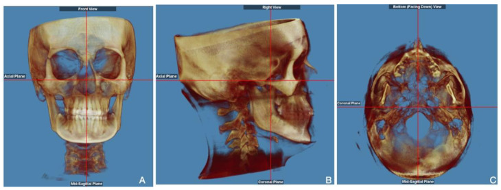
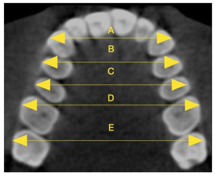
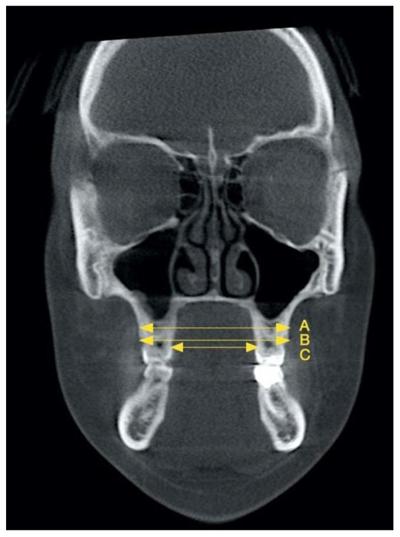
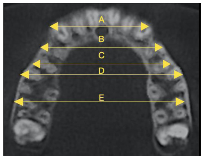
Currently, clear aligners are preferred to conventional appliances, especially among adult patients. However, the use of aligners for treating maxillary constriction is still debated in the literature. Therefore, the purpose of this study was to assess maxillary dentoalveolar expansion following clear aligner therapy in adults using CBCT scans. : The study sample encompassed 50 non-growing patients (27 females and 23 males) aged 20 to 42 undergoing clear aligner orthodontics without dental extractions or auxiliaries. Transverse linear distances were measured on initial and final CBCTs and, subsequently, analysed through paired -test and ANOVA. We considered alveolar bone measurements and interdental widths measured at the buccal apices and cusps from canines to second molars. : The buccal alveolar ridge width showed the greatest expansion (1.01 ± 0.38 mm), followed by the palatal alveolar ridge and maxillary alveolar bone. Statistically significant improvements were observed for all interdental measurements. The most considerable changes occurred in the interpremolar cusp distances, while the least changes were seen in the intermolar apex distances. At the cusp level, the average interpremolar widths increased by 3.44 ± 0.22 mm for the first premolars and 3.14 ± 0.27 mm for the second ones. : Clear aligner treatment can effectively manage a constricted maxillary arch. We found significant changes in the maxillary alveolar bone. Both inter-apex and inter-cusp widths increased in all teeth, with the highest values in the premolars. Moreover, the increases in interdental distances at both apex and cusp levels were related to tooth position.
目前,与传统矫治器相比,透明矫治器更受青睐,尤其是在成年患者中。然而,文献中对于使用透明矫治器治疗上颌缩窄仍存在争议。因此,本研究的目的是通过锥形束计算机断层扫描(CBCT)评估成年患者接受透明矫治器治疗后的上颌牙牙槽扩张情况。:研究样本包括50名年龄在20至42岁之间未生长发育的患者(27名女性和23名男性),他们正在接受透明矫治器正畸治疗,未进行拔牙或使用辅助装置。在初始和最终的CBCT上测量横向线性距离,随后通过配对t检验和方差分析进行分析。我们考虑了牙槽骨测量以及从尖牙到第二磨牙颊尖和牙尖处的牙间宽度。:颊侧牙槽嵴宽度扩张最大(1.01±0.38毫米),其次是腭侧牙槽嵴和上颌牙槽骨。所有牙间测量均观察到具有统计学意义的改善。最显著的变化发生在前磨牙牙尖间距离,而磨牙根尖间距离变化最小。在牙尖水平,第一前磨牙的平均前磨牙间宽度增加了3.44±0.22毫米,第二前磨牙增加了3.14±0.27毫米。:透明矫治器治疗可以有效矫治上颌缩窄。我们发现上颌牙槽骨有显著变化。所有牙齿的根尖间和牙尖间宽度均增加,前磨牙处增加的值最高。此外,根尖和牙尖水平牙间距离的增加与牙齿位置有关。