Mak Sze Mun, Rawal Bhavin, Benedetti Giulia, Eccles Amy, Price Laura, Breen Karen, Padley Simon P G, Karunanithy Narayan
Department of Radiology, Guy's and St Thomas' NHS Foundation Trust, London, SE1 7EH, United Kingdom.
Cardiovascular Imaging Department, King's College London, St Thomas Campus, London, SE1 7AR, United Kingdom.
BJR Open. 2025 Apr 10;7(1):tzaf005. doi: 10.1093/bjro/tzaf005. eCollection 2025 Jan.
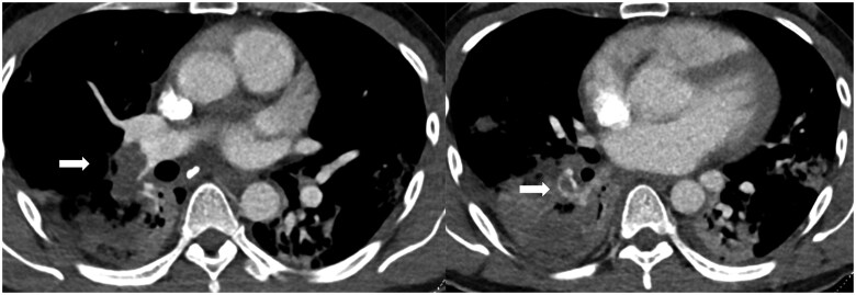
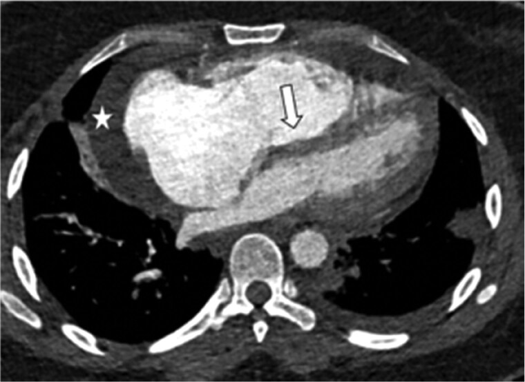
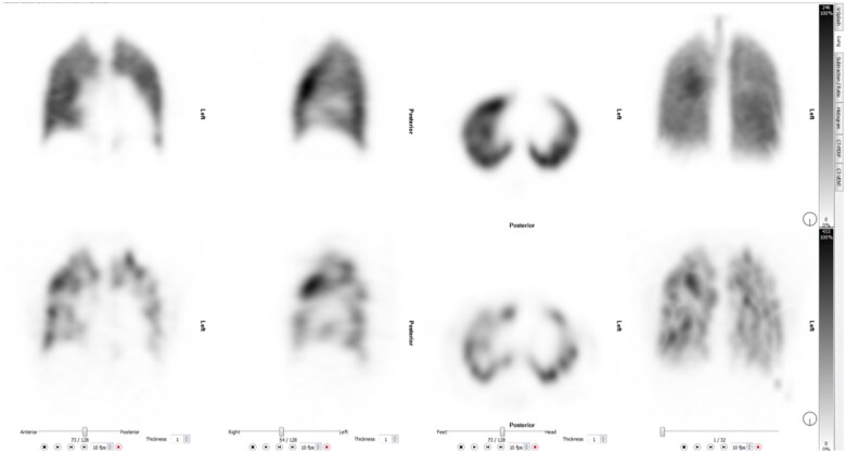
Acute pulmonary emboli can manifest as a spectrum of physiological status ranging from an incidental finding to life threatening right heart failure. We discuss the crucial role imaging plays in the accurate and rapid diagnosis. In addition, imaging features are central in assessing the severity of the presentation allowing for appropriate risk stratification and escalation of care. The relative strengths of the various imaging modalities used in the management of chronic thromboembolic pulmonary hypertension are also discussed.
急性肺栓塞可表现为一系列生理状态,从偶然发现到危及生命的右心衰竭。我们讨论了成像在准确快速诊断中所起的关键作用。此外,成像特征对于评估病情严重程度至关重要,有助于进行适当的风险分层和加强治疗。本文还讨论了用于慢性血栓栓塞性肺动脉高压管理的各种成像方式的相对优势。