Březík Michal, Matušková Veronika, Vysloužilová Daniela, Šín Martin, Chovancová Veronika, Sičová Kristína, Jankaničová Natália, Chrapek Oldřich
Department of Ophthalmology, Faculty of Medicine Masaryk University, Brno, Czech Republic.
Eye Clinic, University Hospital Ostrava, Ostrava, Czech Republic.
Eye Brain. 2025 Jul 8;17:81-94. doi: 10.2147/EB.S524274. eCollection 2025.
Evaluate whether optic disc edema results in changes in retinal microcirculation.
The study group consisted of 11 patients with unilateral optic disc edema (papilledema). The control group consisted of the healthy eyes of the same 11 patients. Patients underwent non-invasive photo-spectrometric retinal oximetry using Oxymap T1 retinal oximeter (Oxymap, Reykjavik, Iceland). In the eyes of these 11 patients, we examined the diameter of the retinal arteries and veins, arterial and venous blood oxygen saturation, and the difference in oxygen saturation between arterioles and venules (A-V difference).
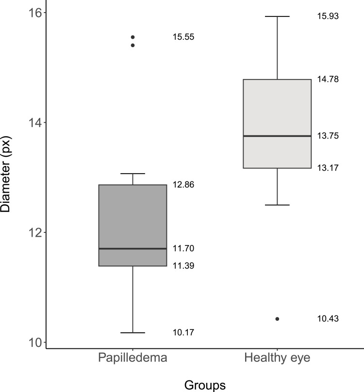
In the papilledema group, the decrease in the retinal arterial diameter was statistically significant (p=0.001). The median diameter of the retinal artery was 11.70 px (IQR 1.47) or after conversion 109.00 µm (IQR 14.00) in the papilledema group and 13.75 px (IQR 1.61) or 128.00 µm (IQR 15.00) in the control group. The increase in the diameter of the retinal veins in the papilledema group was statistically significant (p=0.012), where the median diameter in the papilledema group was 20.88 px (IQR 3.72) or 194.00 µm (IQR 35.00), and in the control group was 18.18 px (IQR 3.60) or 169 µm (IQR 33.00). There was a statistically significant decrease (p<0.001) in the venous saturation in the papilledema group with a median value of 53.16% (IQR 17.38) and 60.02% (IQR 11.98) in the control group. The median of the A-V difference was 51.92 (IQR 15.96) in the papilledema group, resp. 38.49 (IQR 9.75) in the control group and a significant increase in the papilledema group (p<0.001) was reported.
Automatic retinal oximetry demonstrated changes in the retinal microcirculation in patients with optic disc edema.
评估视盘水肿是否会导致视网膜微循环的变化。
研究组由11名单侧视盘水肿(视乳头水肿)患者组成。对照组由这11名患者的健眼组成。患者使用Oxymap T1视网膜血氧仪(Oxymap,冰岛雷克雅未克)接受非侵入性光谱视网膜血氧测定。在这11名患者的眼睛中,我们检测了视网膜动脉和静脉的直径、动脉和静脉血氧饱和度以及小动脉和小静脉之间的血氧饱和度差异(动静脉差异)。
在视乳头水肿组中,视网膜动脉直径的减小具有统计学意义(p = 0.001)。视乳头水肿组视网膜动脉的中位数直径为11.70像素(四分位距1.47),换算后为109.00微米(四分位距14.00),对照组为13.75像素(四分位距1.61)或128.00微米(四分位距15.00)。视乳头水肿组视网膜静脉直径的增加具有统计学意义(p = 0.012),视乳头水肿组的中位数直径为20.88像素(四分位距3.72)或194.00微米(四分位距35.00),对照组为18.18像素(四分位距3.60)或169微米(四分位距33.00)。视乳头水肿组的静脉饱和度有统计学意义的降低(p < 0.001),中位数为53.16%(四分位距17.38),对照组为60.02%(四分位距11.98)。视乳头水肿组的动静脉差异中位数为51.92(四分位距15.96),对照组为38.49(四分位距9.75),且视乳头水肿组有显著增加(p < 0.001)。
自动视网膜血氧测定显示视盘水肿患者的视网膜微循环有变化。