Department of Ophthalmology, University of Southern California, Los Angeles, California, United States.
Richard and Loan Hill Department of Bioengineering, University of Illinois at Chicago, Chicago, Illinois, United States.
Invest Ophthalmol Vis Sci. 2018 Apr 1;59(5):1905-1909. doi: 10.1167/iovs.17-23647.
Reduction in inner retinal oxygen delivery (DO2) can cause retinal hypoxia and impair inner retinal oxygen metabolism (MO2), leading to vision loss. The purpose of the current study was to establish measurements of DO2 and MO2 in healthy subjects and test the hypothesis that DO2 and MO2 are reduced in sickle cell retinopathy (SCR) subjects.
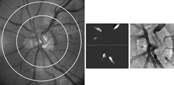
Dual wavelength retinal oximetry and Doppler optical coherence tomography were performed in 12 healthy control and 12 SCR subjects. Images were analyzed to measure retinal arterial and venous oxygen content (O2A and O2V), venous diameter (DV), and total retinal blood flow (TRBF). Retinal arteriovenous oxygen content difference (O2AV), DO2, MO2, and oxygen extraction fraction (OEF) were calculated according to the following equations: O2AV = O2A - O2V; DO2 = TRBF * O2A; MO2 = TRBF * O2AV; OEF = MO2/DO2.
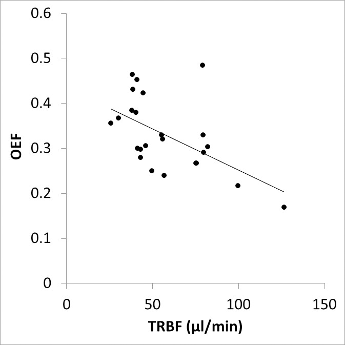
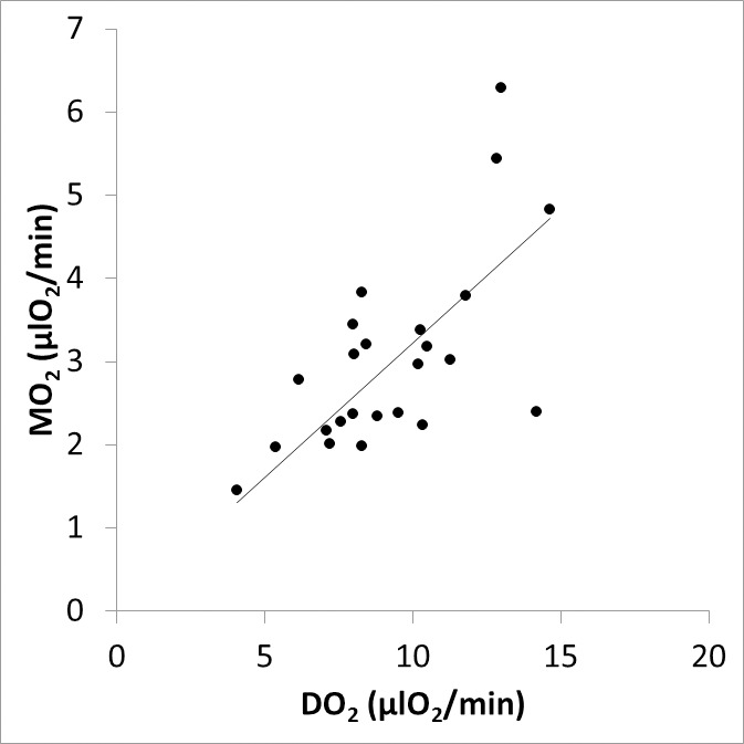
Retinal DV and TRBF were higher in the SCR group as compared to the control group, whereas, O2A, O2V, and O2AV were lower in SCR group as compared to the control group. DO2, MO2, and OEF were not significantly different between control and SCR groups. MO2 and DO2 were linearly related, such that higher MO2 was associated with higher DO2. There was an inverse relationship between TRBF and OEF, such that lower TRBF was associated with higher OEF.
Increased blood flow compensated for decreased oxygen content, thereby maintaining DO2, MO2, and OEF at predominately lower stages of SCR. Quantitative assessment of these parameters has the potential to advance knowledge and improve diagnostic evaluation of retinal ischemic conditions.
视网膜内层氧供减少(DO2)可导致视网膜缺氧,并损害内层视网膜氧代谢(MO2),从而导致视力丧失。本研究的目的是建立健康受试者的 DO2 和 MO2 测量方法,并验证 DO2 和 MO2 在镰状细胞视网膜病变(SCR)患者中降低的假设。
对 12 名健康对照者和 12 名 SCR 患者进行双波长视网膜血氧测定和多普勒光学相干断层扫描。对图像进行分析,以测量视网膜动脉和静脉氧含量(O2A 和 O2V)、静脉直径(DV)和总视网膜血流量(TRBF)。根据以下公式计算视网膜动静脉氧含量差(O2AV)、DO2、MO2 和氧提取分数(OEF):O2AV=O2A-O2V;DO2=TRBFO2A;MO2=TRBFO2AV;OEF=MO2/DO2。
与对照组相比,SCR 组的视网膜 DV 和 TRBF 较高,而 SCR 组的 O2A、O2V 和 O2AV 较低。DO2、MO2 和 OEF 在对照组和 SCR 组之间无显著差异。MO2 和 DO2 呈线性相关,即较高的 MO2 与较高的 DO2 相关。TRBF 与 OEF 呈反比关系,即较低的 TRBF 与较高的 OEF 相关。
血流量增加代偿了氧含量的降低,从而使 DO2、MO2 和 OEF 在 SCR 的主要较低阶段保持不变。这些参数的定量评估有可能提高对视网膜缺血状态的认识和改善诊断评估。