See Cheryl R Z, Si Shuqing, Baird C Lexi, Haswell Courtney C, Hussain Ahmed, Olff Miranda, Veltman Dick J, Frijling Jessie L, van Zuiden Mirjam, Koch Saskia B J, Nawijn Laura, Wang Li, Zhu Ye, Li Gen, Neria Yuval, Zhu Xi, Suarez-Jimenez Benjamin, Zilcha-Mano Sigal, Lazarov Amit, Stevens Jennifer S, Ressler Kerry, Fani Negar, Jovanovic Tanja, van Rooij Sanne J H, Kaufman Milissa L, Lebois Lauren A M, Rosso Isabelle M, Olson Elizabeth A, Baker Justin T, Sponheim Scott R, Disner Seth G, Davenport Nicholas D, Etkin Amit, Maron-Katz Adi, Stein Murray B, Shenton Martha E, Stein Dan J, Ipser Jonathan, Koopowitz Sheri-Michelle, Seedat Soraya, du Plessis Stefan, van den Heuvel Leigh L, Lissek Shmuel, Berg Hannah, Straube Thomas, Hofman David, Baugh Lee A, Forster Gina L, Simons Raluca M, Simons Jeffrey S, Magnotta Vincent A, Fercho Kelene A, Wang Xin, Cotton Andrew S, O'Leary Erin N, Xie Hong, Grupe Daniel W, Nitschke Jack B, Davidson Richard J, Larson Christine L, deRoon-Cassini Terri A, Tomas Carissa W, Fitzgerald Jacklynn M, Blackford Jennifer Urbano, Olatunji Bunmi O, Gordon Evan M, May Geoffrey, Nelson Steven M, Lanius Ruth, Théberge Jean, Densmore Maria, Neufeld Richard W J, Abdallah Chadi G, Averill Christopher L, Harpaz-Rotem Ilan, Levy Ifat, Krystal John H, Geuze Elbert, van Lutterveld Remko, Dennis Emily L, Tate David F, Cifu David X, Walker William C, Wilde Elisabeth A, van der Wee Nic J A, Vermeiren Robert R J M, van der Werff Steven J A, McLaughlin Katie, Sambrook Kelly, Peverill Matthew, Radua Joaquim, Salminen Lauren E, Jahanshad Neda, Thomopoulos Sophia I, James Anthony, Valmaggia Lucia, Thompson Paul M, Morey Rajendra A, Kempton Matthew J
Department of Psychosis, Institute of Psychiatry, Psychology & Neuroscience, King's College London, London, UK.
Brain Imaging and Analysis Center, Duke University, Durham, NC, USA.
Eur Psychiatry. 2025 Jul 22;68(1):e97. doi: 10.1192/j.eurpsy.2025.10062.
Patients with posttraumatic stress disorder (PTSD) exhibit smaller regional brain volumes in commonly reported regions including the amygdala and hippocampus, regions associated with fear and memory processing. In the current study, we have conducted a voxel-based morphometry (VBM) meta-analysis using whole-brain statistical maps with neuroimaging data from the ENIGMA-PGC PTSD working group.
T1-weighted structural neuroimaging scans from 36 cohorts (PTSD = 1309; controls = 2198) were processed using a standardized VBM pipeline (ENIGMA-VBM tool). We meta-analyzed the resulting statistical maps for voxel-wise differences in gray matter (GM) and white matter (WM) volumes between PTSD patients and controls, performed subgroup analyses considering the trauma exposure of the controls, and examined associations between regional brain volumes and clinical variables including PTSD (CAPS-4/5, PCL-5) and depression severity (BDI-II, PHQ-9).
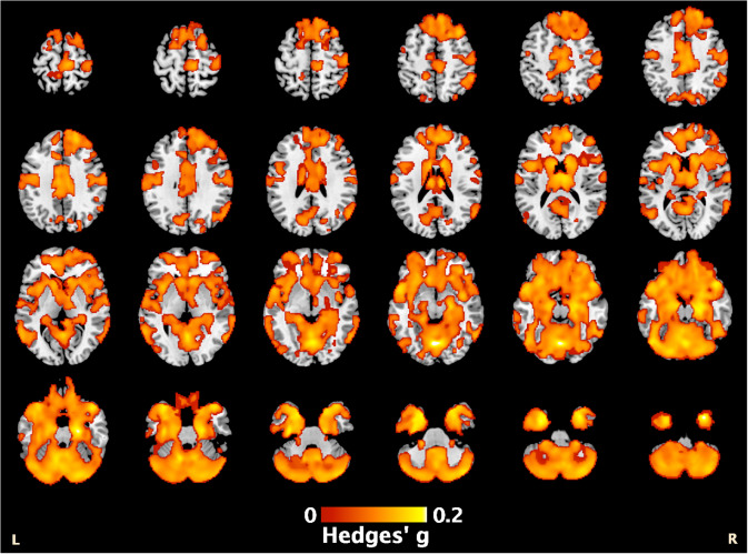
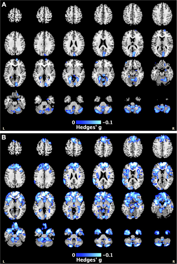
PTSD patients exhibited smaller GM volumes across the frontal and temporal lobes, and cerebellum, with the most significant effect in the left cerebellum (Hedges' = 0.22, = .001), and smaller cerebellar WM volume (peak Hedges' = 0.14, = .008). We observed similar regional differences when comparing patients to trauma-exposed controls, suggesting these structural abnormalities may be specific to PTSD. Regression analyses revealed PTSD severity was negatively associated with GM volumes within the cerebellum ( = .003), while depression severity was negatively associated with GM volumes within the cerebellum and superior frontal gyrus in patients ( = .001).
PTSD patients exhibited widespread, regional differences in brain volumes where greater regional deficits appeared to reflect more severe symptoms. Our findings add to the growing literature implicating the cerebellum in PTSD psychopathology.
创伤后应激障碍(PTSD)患者在包括杏仁核和海马体等常见报道区域的脑区体积较小,这些区域与恐惧和记忆处理相关。在本研究中,我们使用来自ENIGMA-PGC PTSD工作组的神经影像数据,通过全脑统计图进行了基于体素的形态学(VBM)荟萃分析。
使用标准化的VBM流程(ENIGMA-VBM工具)处理来自36个队列(PTSD患者 = 1309例;对照组 = 2198例)的T1加权结构神经影像扫描。我们对PTSD患者和对照组之间灰质(GM)和白质(WM)体积的体素差异的统计地图进行荟萃分析,考虑对照组的创伤暴露情况进行亚组分析,并检查脑区体积与临床变量之间的关联,包括PTSD(CAPS-4/5,PCL-5)和抑郁严重程度(BDI-II,PHQ-9)。
PTSD患者在额叶、颞叶和小脑的GM体积较小,在左小脑的影响最为显著(Hedges' = −0.22,P = .001),小脑WM体积也较小(峰值Hedges' = −0.14,P = .008)。将患者与有创伤暴露的对照组进行比较时,我们观察到了类似的区域差异,表明这些结构异常可能是PTSD特有的。回归分析显示,PTSD严重程度与小脑中的GM体积呈负相关(P = .003),而抑郁严重程度与患者小脑中的GM体积以及额上回的GM体积呈负相关(P = .001)。
PTSD患者在脑体积上表现出广泛的区域差异,其中更大的区域缺陷似乎反映了更严重的症状。我们的研究结果进一步丰富了越来越多的将小脑与PTSD精神病理学联系起来的文献。