• 文献检索
  • 文档翻译
  • 深度研究
  • 学术资讯
  • Suppr Zotero 插件Zotero 插件
  • 邀请有礼
  • 套餐&价格
  • 历史记录
应用&插件
Suppr Zotero 插件Zotero 插件浏览器插件Mac 客户端Windows 客户端微信小程序
定价
高级版会员购买积分包购买API积分包
服务
文献检索文档翻译深度研究API 文档MCP 服务
关于我们
关于 Suppr公司介绍联系我们用户协议隐私条款
关注我们

Suppr 超能文献

核心技术专利:CN118964589B侵权必究
粤ICP备2023148730 号-1Suppr @ 2026

文献检索

告别复杂PubMed语法,用中文像聊天一样搜索,搜遍4000万医学文献。AI智能推荐,让科研检索更轻松。

立即免费搜索

文件翻译

保留排版,准确专业,支持PDF/Word/PPT等文件格式,支持 12+语言互译。

免费翻译文档

深度研究

AI帮你快速写综述,25分钟生成高质量综述,智能提取关键信息,辅助科研写作。

立即免费体验

人中央凹小凹的体内视锥光感受器地形图

In Vivo Cone Photoreceptor Topography of the Human Foveola.

作者信息

Ameln Julius, Witten Jenny L, Gutnikov Aleksandr, Lukyanova Veronika, Holz Frank G, Harmening Wolf M

机构信息

Department of Ophthalmology, University of Bonn, Bonn, Germany.

出版信息

Invest Ophthalmol Vis Sci. 2025 Aug 1;66(11):13. doi: 10.1167/iovs.66.11.13.

DOI:10.1167/iovs.66.11.13
PMID:40767443
原文链接:https://pmc.ncbi.nlm.nih.gov/articles/PMC12347159/
Abstract

PURPOSE

To study in vivo cone topography of the normal human foveola.

METHODS

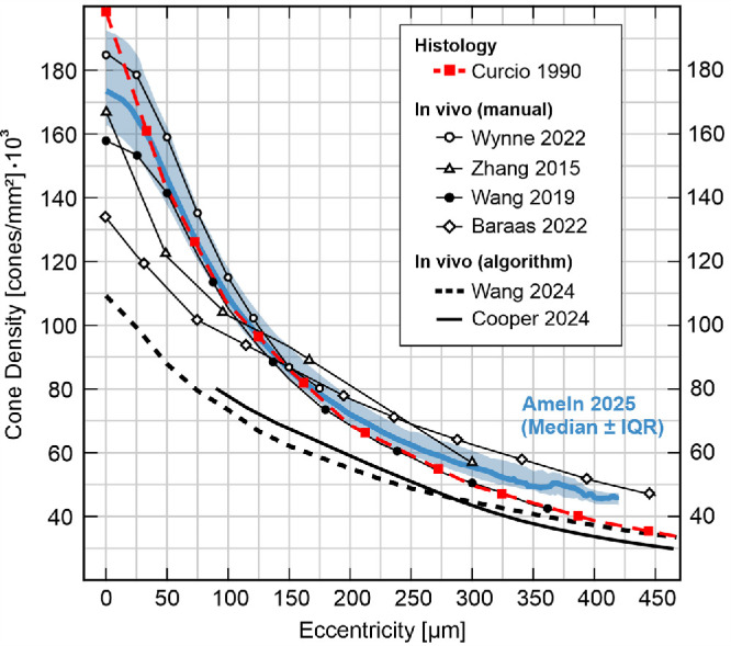
The fovea in both eyes of 30 healthy participants was imaged with adaptive optics scanning light ophthalmoscopy. High-resolution image montages spanning two degrees of visual angle were created and cone center locations annotated. Continuous cone density maps were computed by a Voronoi cell area approach to also yield the topographical center, the cone density centroid (CDC). Cone density profiles were extracted and fit with a four-parameter decay function, D = D0 / (1 + (E/a)b)c, with D as cone density (cones/mm2), D0 as cone density at the CDC, and E as eccentricity (µm).

RESULTS

Across eyes, D0 was 175,474 ± 20,543 cones/mm2, on average (range 136,001-216,209 cones/mm2). Density dropped anisotropically along the meridians, shallower horizontally, with average best fit parameters (a, b, c) of 61.95, 2.469, 0.268 for horizontal, and 59.11, 2.012, 0.357, for vertical profiles, respectively. In radially averaged profiles, cone density reached 50% of D0 at 151 ± 17 µm eccentricity (range 128-193 µm). Temporal cone density was slightly higher than nasal. Most topographical metrics were highly correlated between fellow eyes.

CONCLUSIONS

Despite a 1.6-fold range in absolute cone density, foveolar density profiles could be well described by a sigmoidal decay function across all eyes. This established a normative cone density profile of the healthy foveola. It allowed cone density estimation in cases of only partially available data, which alleviates resolution demands for future studies and renders possible retrospective analyses of foveolar cone topography in sub-optimal imagery.

摘要

目的

研究正常人中心小凹的体内视锥细胞地形图。

方法

使用自适应光学扫描激光检眼镜对30名健康受试者双眼的中央凹进行成像。创建跨越两个视角的高分辨率图像蒙太奇,并标注视锥细胞中心位置。通过Voronoi细胞面积法计算连续的视锥细胞密度图,以得出地形中心,即视锥细胞密度质心(CDC)。提取视锥细胞密度分布曲线,并用四参数衰减函数D = D0 / (1 + (E/a)b)c进行拟合,其中D为视锥细胞密度(个/mm2),D0为CDC处的视锥细胞密度,E为偏心率(μm)。

结果

双眼的D0平均为175,474 ± 20,543个/mm2(范围为136,001 - 216,209个/mm2)。密度沿子午线各向异性下降,水平方向下降较浅,水平分布曲线的平均最佳拟合参数(a、b、c)分别为61.95、2.469、0.268,垂直分布曲线的平均最佳拟合参数分别为59.11、2.012、0.357。在径向平均分布曲线中,视锥细胞密度在偏心率为151 ± 17 µm(范围为128 - 193 µm)时达到D0的50%。颞侧视锥细胞密度略高于鼻侧。大多数地形学指标在双眼之间高度相关。

结论

尽管视锥细胞绝对密度范围为1.6倍,但所有眼睛的中心小凹密度分布曲线都可以用S形衰减函数很好地描述。这建立了健康中心小凹的标准视锥细胞密度分布。它允许在仅有部分可用数据的情况下估计视锥细胞密度,这减轻了未来研究的分辨率要求,并使得对次优图像中的中心小凹视锥细胞地形图进行回顾性分析成为可能。

https://cdn.ncbi.nlm.nih.gov/pmc/blobs/3b56/12347159/406ba6abac85/iovs-66-11-13-f008.jpg
https://cdn.ncbi.nlm.nih.gov/pmc/blobs/3b56/12347159/d087c7c33e04/iovs-66-11-13-f001.jpg
https://cdn.ncbi.nlm.nih.gov/pmc/blobs/3b56/12347159/68d0fc966139/iovs-66-11-13-f002.jpg
https://cdn.ncbi.nlm.nih.gov/pmc/blobs/3b56/12347159/47d34d9dbf7d/iovs-66-11-13-f003.jpg
https://cdn.ncbi.nlm.nih.gov/pmc/blobs/3b56/12347159/ded5375dbb4b/iovs-66-11-13-f004.jpg
https://cdn.ncbi.nlm.nih.gov/pmc/blobs/3b56/12347159/0a84ff118e79/iovs-66-11-13-f005.jpg
https://cdn.ncbi.nlm.nih.gov/pmc/blobs/3b56/12347159/cdbeddd20c9e/iovs-66-11-13-f006.jpg
https://cdn.ncbi.nlm.nih.gov/pmc/blobs/3b56/12347159/1fc158531c90/iovs-66-11-13-f007.jpg
https://cdn.ncbi.nlm.nih.gov/pmc/blobs/3b56/12347159/406ba6abac85/iovs-66-11-13-f008.jpg

相似文献

1
In Vivo Cone Photoreceptor Topography of the Human Foveola.人中央凹小凹的体内视锥光感受器地形图
Invest Ophthalmol Vis Sci. 2025 Aug 1;66(11):13. doi: 10.1167/iovs.66.11.13.
2
Cone Density Changes After Repeated Low-Level Red Light Treatment in Children With Myopia.近视儿童反复接受低强度红光治疗后视锥细胞密度变化
JAMA Ophthalmol. 2025 Apr 24. doi: 10.1001/jamaophthalmol.2025.0835.
3
Intervisit Reproducibility of Foveal Cone Density Metrics.黄斑中心凹锥体细胞密度指标的随访可重复性。
Transl Vis Sci Technol. 2024 Jun 3;13(6):18. doi: 10.1167/tvst.13.6.18.
4
Multimodal High-Resolution Imaging in Retinitis Pigmentosa: A Comparison Between Optoretinography, Cone Density, and Visual Sensitivity.多模态高分辨率成像在色素性视网膜炎中的应用:光感受器图、锥体细胞密度和视觉敏感性的比较。
Invest Ophthalmol Vis Sci. 2024 Aug 1;65(10):45. doi: 10.1167/iovs.65.10.45.
5
LONGITUDINAL ADAPTIVE OPTICS SCANNING LASER OPHTHALMOSCOPY REVEALS REGIONAL VARIATION IN CONE AND ROD PHOTORECEPTOR LOSS IN STARGARDT DISEASE.纵向自适应光学扫描激光检眼镜显示斯塔加特病中视锥和视杆光感受器的丢失存在区域性差异。
Retina. 2024 Aug 1;44(8):1403-1412. doi: 10.1097/IAE.0000000000004104.
6
Variability in Human Cone Topography Assessed by Adaptive Optics Scanning Laser Ophthalmoscopy.通过自适应光学扫描激光眼科显微镜评估的人类视锥细胞地形图的变异性。
Am J Ophthalmol. 2015 Aug;160(2):290-300.e1. doi: 10.1016/j.ajo.2015.04.034. Epub 2015 Apr 30.
7
Prescription of Controlled Substances: Benefits and Risks管制药品的处方:益处与风险
8
The Relationship Between Visual Sensitivity and Eccentricity, Cone Density and Outer Segment Length in the Human Foveola.人中央凹的视觉敏感度与离轴距离、视锥细胞密度和外节长度的关系。
Invest Ophthalmol Vis Sci. 2021 Jul 1;62(9):31. doi: 10.1167/iovs.62.9.31.
9
Adaptive-optics scanning laser ophthalmoscopy study of non-arteritic anterior ischemic optic neuropathy.非动脉炎性前部缺血性视神经病变的自适应光学扫描激光检眼镜研究
Photodiagnosis Photodyn Ther. 2025 Aug;54:104678. doi: 10.1016/j.pdpdt.2025.104678. Epub 2025 Jun 13.
10
Retinal Vascular Diseases Highlighted by Adaptive Optics Ophthalmoscopy.自适应光学检眼镜凸显的视网膜血管疾病
Rom J Ophthalmol. 2025 Apr-Jun;69(2):235-242. doi: 10.22336/rjo.2025.37.

引用本文的文献

1
Neural network assisted annotation and analysis tool to study in-vivo foveolar cone photoreceptor topography.用于研究体内小凹锥状光感受器地形图的神经网络辅助注释和分析工具。
Sci Rep. 2025 Jul 4;15(1):23858. doi: 10.1038/s41598-025-08028-9.

本文引用的文献

1
Neural network assisted annotation and analysis tool to study in-vivo foveolar cone photoreceptor topography.用于研究体内小凹锥状光感受器地形图的神经网络辅助注释和分析工具。
Sci Rep. 2025 Jul 4;15(1):23858. doi: 10.1038/s41598-025-08028-9.
2
Advances in Electrode Design and Physiological Considerations for Retinal Implants.视网膜植入物的电极设计进展及生理考量
Micromachines (Basel). 2025 May 21;16(5):598. doi: 10.3390/mi16050598.
3
Voretigene neparvovec in RPE65-related inherited retinal dystrophy: the 1-year real-world study LIGHT.
用于RPE65相关遗传性视网膜营养不良的voretigene neparvovec:1年真实世界研究LIGHT
Eye (Lond). 2025 Jun;39(9):1758-1764. doi: 10.1038/s41433-025-03691-8. Epub 2025 Mar 14.
4
Evaluation of Retinal Sensitivity in Complete Retinal-Pigment-Epithelium and Outer Retinal Atrophy (cRORA) Lesions in Intermediate Age-Related Macular Degeneration (iAMD) by High-Resolution Microperimetry.通过高分辨率微视野计评估中度年龄相关性黄斑变性(iAMD)中完全视网膜色素上皮和外层视网膜萎缩(cRORA)病变的视网膜敏感性
J Clin Med. 2024 Dec 20;13(24):7785. doi: 10.3390/jcm13247785.
5
Stem cell-based therapies for retinal diseases: focus on clinical trials and future prospects.基于干细胞的视网膜疾病治疗:聚焦于临床试验与未来前景。
Ophthalmic Genet. 2024 Nov 15:1-14. doi: 10.1080/13816810.2024.2423784.
6
Sub-cone visual resolution by active, adaptive sampling in the human foveola.人中央凹的主动、自适应采样的亚锥视分辨率。
Elife. 2024 Oct 29;13:RP98648. doi: 10.7554/eLife.98648.
7
Morphology of the normative human cone photoreceptor mosaic and a publicly available adaptive optics montage repository.正常人类视锥细胞光感受器嵌合体形态及公开自适应光学拼接库。
Sci Rep. 2024 Oct 5;14(1):23166. doi: 10.1038/s41598-024-74274-y.
8
Gene therapy for retinal diseases: From genetics to treatment.基因治疗视网膜疾病:从遗传学角度到治疗方法。
Indian J Ophthalmol. 2024 Aug 1;72(8):1091-1101. doi: 10.4103/IJO.IJO_2902_23. Epub 2024 Jul 29.
9
The effect of sampling window size on topographical maps of foveal cone density.采样窗口大小对中央凹视锥细胞密度地形图的影响。
Front Ophthalmol (Lausanne). 2024 Apr 9;4:1348950. doi: 10.3389/fopht.2024.1348950. eCollection 2024.
10
Assessment of local sensitivity in incomplete retinal pigment epithelium and outer retinal atrophy (iRORA) lesions in intermediate age-related macular degeneration (iAMD).评估中年相关年龄相关性黄斑变性(iAMD)中不完全性视网膜色素上皮和外层视网膜萎缩(iRORA)病变的局部敏感性。
BMJ Open Ophthalmol. 2024 Jul 9;9(1):e001638. doi: 10.1136/bmjophth-2024-001638.