Ohashi Masayuki, Hasegawa Kazuhiro, Hatsushikano Shun, Imai Norio, Tashi Hideki, Shibuya Yohei, Minato Keitaro, Sato Masayuki, Sekimoto Hiroyuki, Watanabe Kei, Kawashima Hiroyuki
Division of Orthopedic Surgery, Department of Regenerative and Transplant Medicine, Niigata University Graduate School of Medical and Dental Sciences, Niigata, Japan.
Niigata Spine Surgery Center, Niigata, Japan.
Spine Surg Relat Res. 2024 Dec 20;9(4):469-476. doi: 10.22603/ssrr.2024-0283. eCollection 2025 Jul 27.
To estimate natural standing sagittal alignment in patients with adult spinal deformity (ASD), we previously reported the normative values of anatomical pelvic parameters in a healthy population, based on the anterior pelvic plane (APP), and observed the relationships between anatomical and positional pelvic parameters in the standing position. As the second step, we aim to investigate the relationships between anatomical pelvic parameters and standing spinal sagittal alignment in a healthy population.
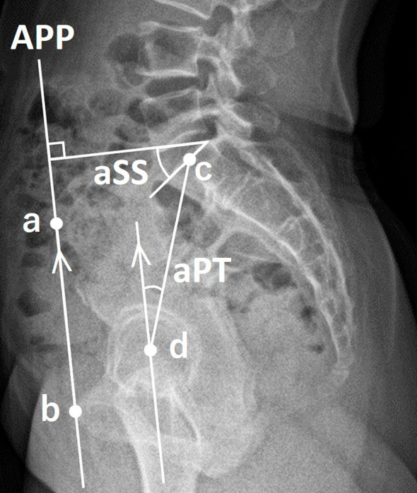
We analyzed biplanar, slot-scanning, full-body stereo radiography of 140 healthy Japanese volunteers (mean age, 39.5 years; 59.3% women). The APP was defined by bilateral anterior superior iliac spines and anterior surface of the pubis symphysis. Anatomical sacral slope (aSS) and anatomical pelvic tilt (aPT) were calculated as angles of the SS and PT regarding the APP.
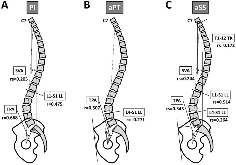
The APP was tilted anteriorly in the sagittal plane by an average of 0.7°. Anatomical pelvic parameters significantly correlated with standing sagittal parameters, except for cervical lordosis and T4-12 thoracic kyphosis (TK) (p<0.05). L4-S1 lumbar lordosis (LL) significantly correlated with aPT and aSS, but not with pelvic incidence (PI). In addition, T1-12 TK significantly correlated with aSS. Multiple linear regression analysis for lumbar alignment produced the following equations: L1-S1 LL (°)=0.588×aSS+30.522, L4-S1 LL (°)=0.165×aSS-0.248×aPT+32.825, lordosis distribution index (%)=-0.662×PI+102.8.
Novel relationships in a healthy population were identified between the anatomical characteristics of the pelvis and standing sagittal parameters not represented by PI. This novel measurement concept based on the APP may estimate natural standing sagittal alignments and proportions using anatomical pelvic parameters in ASD.
为了评估成人脊柱畸形(ASD)患者的自然站立矢状面排列情况,我们之前报告了基于骨盆前平面(APP)的健康人群解剖学骨盆参数的正常值,并观察了站立位时解剖学和位置性骨盆参数之间的关系。作为第二步,我们旨在研究健康人群中解剖学骨盆参数与站立位脊柱矢状面排列之间的关系。
我们分析了140名健康日本志愿者(平均年龄39.5岁;59.3%为女性)的双平面、缝隙扫描全身立体X线摄影。APP由双侧髂前上棘和耻骨联合前表面定义。解剖学骶骨倾斜角(aSS)和解剖学骨盆倾斜角(aPT)计算为骶骨倾斜角(SS)和骨盆倾斜角(PT)相对于APP的角度。
APP在矢状面平均向前倾斜0.7°。除颈椎前凸和T4 - 12胸椎后凸(TK)外,解剖学骨盆参数与站立矢状面参数显著相关(p<0.05)。L4 - S1腰椎前凸(LL)与aPT和aSS显著相关,但与骨盆入射角(PI)无关。此外,T1 - 12 TK与aSS显著相关。腰椎排列的多元线性回归分析得出以下方程:L1 - S1 LL(°)=0.588×aSS + 30.522,L4 - S1 LL(°)=0.165×aSS - 0.248×aPT + 32.825,前凸分布指数(%)=-0.662×PI + 102.8。
在健康人群中发现了骨盆解剖学特征与非PI所代表的站立矢状面参数之间的新关系。这种基于APP的新测量概念可能使用ASD患者的解剖学骨盆参数来估计自然站立矢状面排列和比例。