Ohshima Shusuke, Quispe-Salcedo Angela, Ida-Yonemochi Hiroko, Ueki Yushi, Horii Arata, Ohshima Hayato
Department of Otolaryngology Head and Neck Surgery, Niigata University Graduate School of Medical and Dental Sciences, Niigata, Japan.
Division of Anatomy and Cell Biology of the Hard Tissue, Department of Tissue Regeneration and Reconstruction, Niigata University Graduate School of Medical and Dental Sciences, Niigata, Japan.
Regen Ther. 2025 Aug 13;30:595-605. doi: 10.1016/j.reth.2025.08.007. eCollection 2025 Dec.
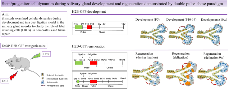
Active and quiescent stem cells coexist in hair follicles and intestinal crypts; however, their localization and differentiation potential in the salivary dynamics are unknown. This study aimed to clarify the cellular dynamics that occur during salivary gland development and regeneration in the duct ligation model with a focus on the role of label retaining cells (LRCs), presumably quiescent stem cells, in these processes.
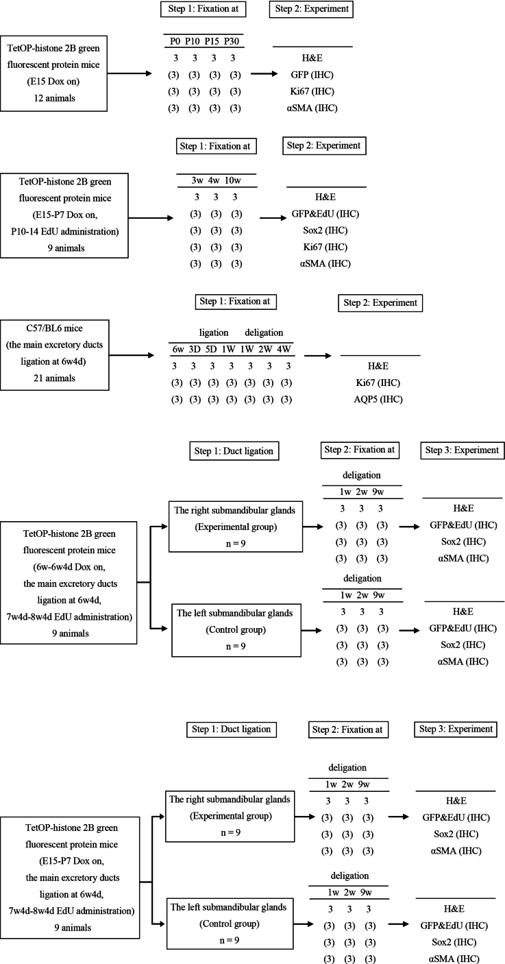
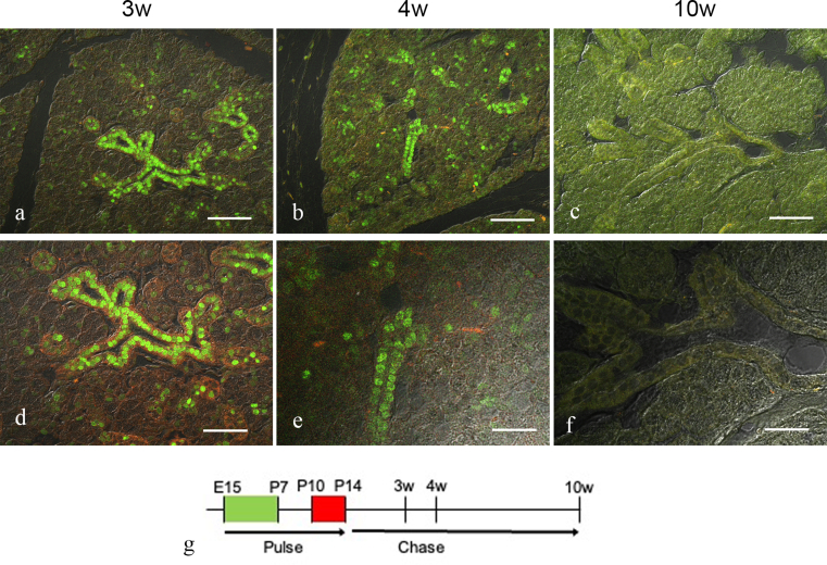
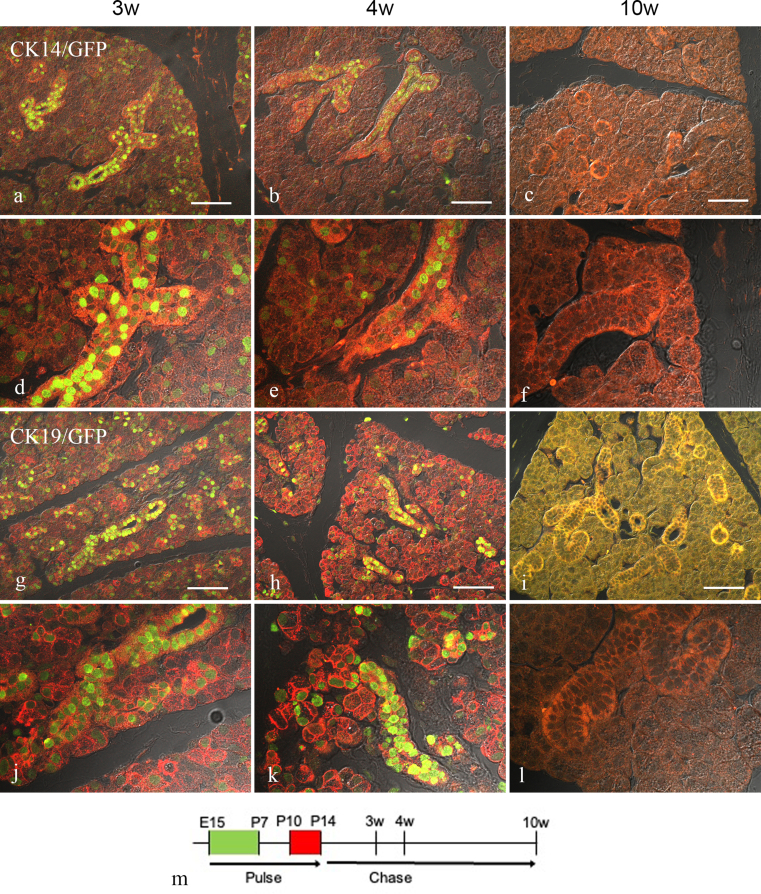
Doxycycline-inducible TetOP-histone 2B (H2B)-green fluorescent protein (GFP) transgenic mice [GFP expression was induced during embryonic day 15 (E15)-postnatal day 7 (P7)] followed by EdU (5-ethynyl-2'-deoxyuridine) administration at P10-14 to chase the LRCs during development. In addition, LRCs were labeled with GFP immediately before salivary gland duct ligation and EdU was administered after the ligation was released to chase the LRCs with GFP and EdU during tissue repair.
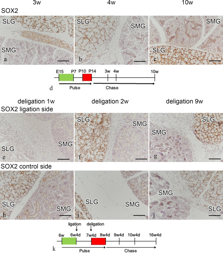
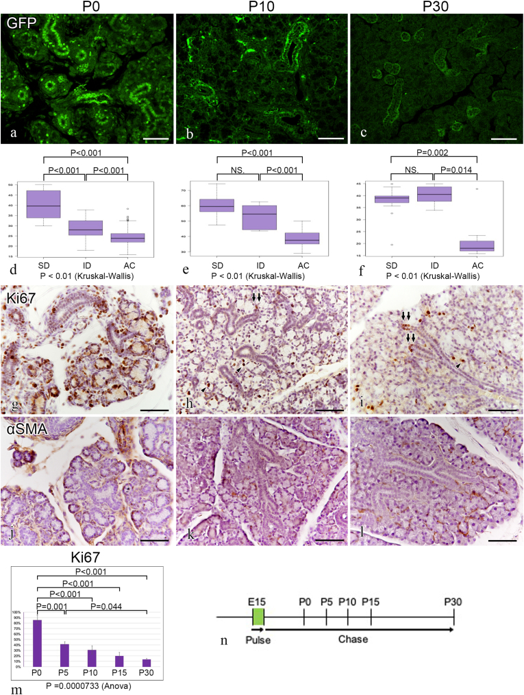
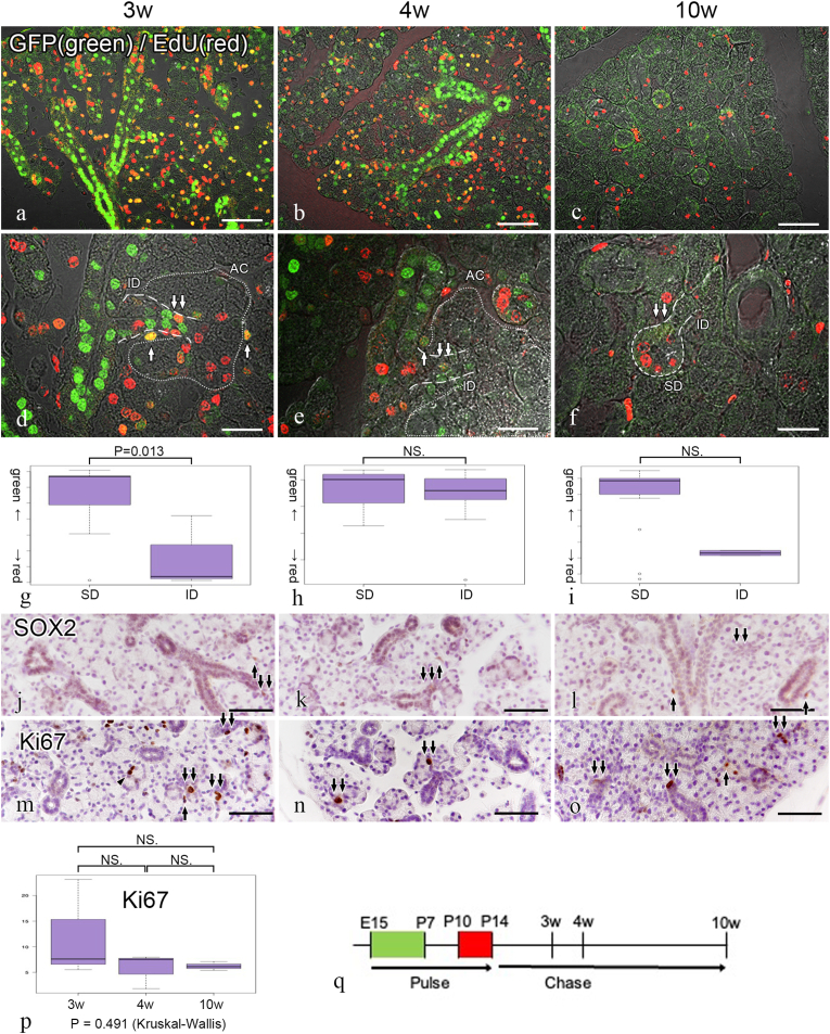
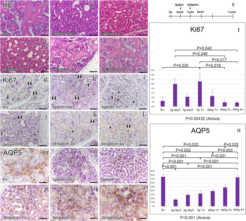
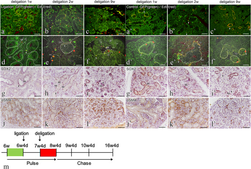
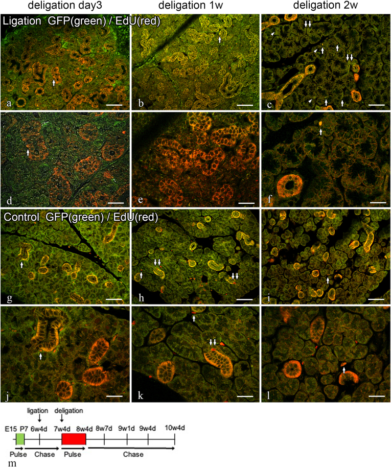
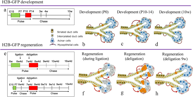
During development, GFP (+) EdU (-) LRCs were abundant in striated duct cells (SDCs) and GFP (+) EdU (+) LRCs were primarily localized to the intercalated duct cells (IDCs) at P21. Labeling of the GFP (+) LRCs faded as well as the EdU (+) LRCs at P70. During tissue repair, GFP (+) EdU (+) LRCs were colocalized in the IDCs and myoepithelium cells (MECs), whereas the GFP (+) EdU (+) acinar cells (ACs) appeared over time. These results suggest that salivary gland quiescent stem/progenitor cells are present in the IDCs during development and that quiescent stem/progenitor cells in the IDCs and MECs differentiate into ACs during tissue repair.
活跃和静止的干细胞共存于毛囊和肠隐窝中;然而,它们在唾液腺动态变化中的定位和分化潜能尚不清楚。本研究旨在阐明在导管结扎模型中唾液腺发育和再生过程中发生的细胞动态变化,重点关注标记保留细胞(LRCs)(可能是静止干细胞)在这些过程中的作用。
用强力霉素诱导的TetOP-组蛋白2B(H2B)-绿色荧光蛋白(GFP)转基因小鼠[在胚胎第15天(E15)至出生后第7天(P7)诱导GFP表达],然后在出生后第10 - 14天给予EdU(5-乙炔基-2'-脱氧尿苷),以追踪发育过程中的LRCs。此外,在唾液腺导管结扎前立即用GFP标记LRCs,并在结扎解除后给予EdU,以在组织修复过程中追踪带有GFP和EdU的LRCs。
在发育过程中,出生后第21天,GFP(+)EdU(-)LRCs在纹状管细胞(SDCs)中丰富,而GFP(+)EdU(+)LRCs主要定位于闰管细胞(IDCs)。在出生后第70天,GFP(+)LRCs以及EdU(+)LRCs的标记消失。在组织修复过程中,GFP(+)EdU(+)LRCs共定位于IDCs和肌上皮细胞(MECs),而GFP(+)EdU(+)腺泡细胞(ACs)随时间出现。这些结果表明,唾液腺静止干细胞/祖细胞在发育过程中存在于IDCs中,并且在组织修复过程中,IDCs和MECs中的静止干细胞/祖细胞分化为ACs。