Bellamy J E, Latshaw W K, Nielsen N O
Can J Comp Med. 1973 Jan;37(1):56-62.
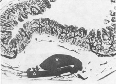
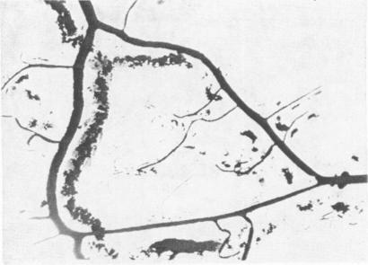
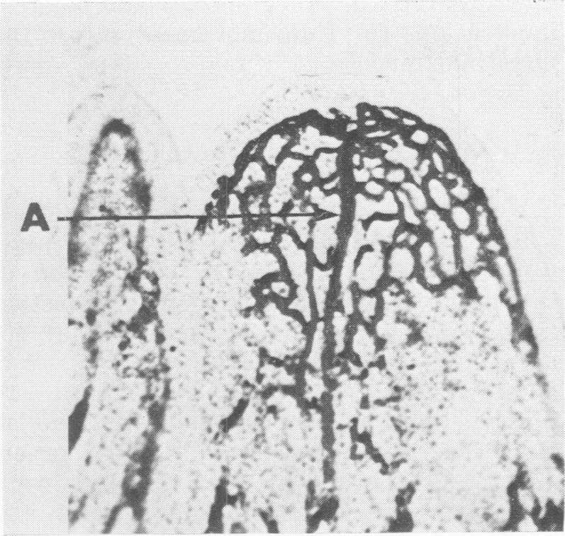
The vascular anatomy of the porcine small intestine was studied by injection of intestinal vessels with India Ink. Examination of transverse and longitudinal serial sections of the injected intestine facilitated a three-dimensional interpretation of the vascular pattern. An artery from the mesentery penetrated the tunica muscularis, supplied muscular branches and passed on to the submucosa where it formed an arterial rete. From the submucosal arteries, arterioles arose and followed a direct axial course to the tips of villi where they ramified into a subepithelial capillary plexus. Some of the capillaries, at the midpoint of the villus, fused into paraxial venules, which emptied into a "transverse venule" at the base of the villus. Other villus capillaries were continuous with those of the crypts. The pericryptal capillary plexus received a few arterial branches from the submucosal arteries. The transverse venule and the pericryptal capillary plexus emptied into large, segmentally dilated veins in the submucosa. The submucosal veins formed an extensive anastomosing network drained by large venous trunks which passed through the muscle layers to the mesentery. The observations suggest possible relationships between the vascular pattern and intestinal fluid movement.
通过向猪小肠血管注射印度墨水来研究其血管解剖结构。对注射后的小肠横切和纵切连续切片进行检查,有助于对血管模式进行三维解读。来自肠系膜的一条动脉穿透肌层,供应肌支,然后进入黏膜下层,在那里形成一个动脉网。黏膜下动脉发出小动脉,沿直接的轴向路径到达绒毛尖端,在那里分支形成上皮下毛细血管丛。一些位于绒毛中点的毛细血管融合成近轴小静脉,这些小静脉在绒毛基部汇入一条“横向小静脉”。其他绒毛毛细血管与隐窝的毛细血管相连。隐窝周围毛细血管丛从黏膜下动脉接收一些动脉分支。横向小静脉和隐窝周围毛细血管丛汇入黏膜下层的大的、节段性扩张的静脉。黏膜下静脉形成广泛的吻合网络,由大的静脉干引流,这些静脉干穿过肌层到达肠系膜。这些观察结果提示了血管模式与肠液流动之间可能存在的关系。