• 文献检索
  • 文档翻译
  • 深度研究
  • 学术资讯
  • Suppr Zotero 插件Zotero 插件
  • 邀请有礼
  • 套餐&价格
  • 历史记录
应用&插件
Suppr Zotero 插件Zotero 插件浏览器插件Mac 客户端Windows 客户端微信小程序
定价
高级版会员购买积分包购买API积分包
服务
文献检索文档翻译深度研究API 文档MCP 服务
关于我们
关于 Suppr公司介绍联系我们用户协议隐私条款
关注我们

Suppr 超能文献

核心技术专利:CN118964589B侵权必究
粤ICP备2023148730 号-1Suppr @ 2026

文献检索

告别复杂PubMed语法,用中文像聊天一样搜索,搜遍4000万医学文献。AI智能推荐,让科研检索更轻松。

立即免费搜索

文件翻译

保留排版,准确专业,支持PDF/Word/PPT等文件格式,支持 12+语言互译。

免费翻译文档

深度研究

AI帮你快速写综述,25分钟生成高质量综述,智能提取关键信息,辅助科研写作。

立即免费体验

细菌膜和细胞壁的电子显微镜研究以及生长起始过程中发生的变化。

Electron microscopic study of membranes and walls of bacteria and changes occurring during growth initiation.

作者信息

Hurst A, Stubbs J M

出版信息

J Bacteriol. 1969 Mar;97(3):1466-79. doi: 10.1128/jb.97.3.1466-1479.1969.

DOI:10.1128/jb.97.3.1466-1479.1969
PMID:4180467
原文链接:https://pmc.ncbi.nlm.nih.gov/articles/PMC249870/
Abstract

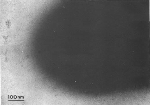
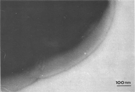
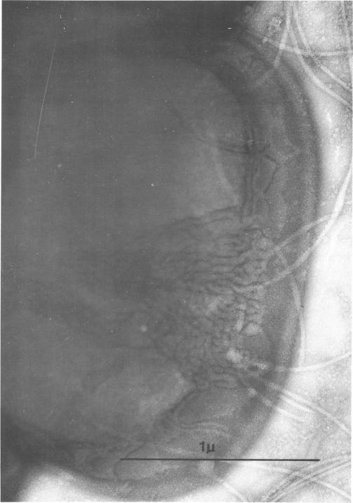
Thin sections of stationary-phase Streptococcus lactis cells showed that the wall and membrane are 20 and 7 nm thick, respectively. Whole cells were examined by negative staining with ammonium molybdate and by shadowing. On air-drying of whole cells, the membrane pulled away from the wall revealing adhesions between these organelles. Adhesions could not be seen after subculture of the stationary-phase cells into complex media or into solutions containing glucose, KCl, and CaCl(2) in tris(hydroxymethyl)aminomethane buffer. The adhesions were also observed in stationary-phase cells of other gram-positive bacteria. Fractured freeze-etched cells of S. lactis had a smooth outside surface, but the inside of the wall (or outside of the membrane) had a regular structure, repeating at 10 nm, which could correspond to the adhesions observed in the negatively stained air-dried cells. Freeze-etching also revealed holes in the outside wall which had the shape of inverted truncated cones. The outside diameter of the cone was 60 nm, and the diameter on the inside surface of the wall was 20 nm. The membrane had upstanding plugs, 20 nm in diameter, which could fill the holes in the wall.

摘要

对处于稳定期的乳酸链球菌细胞进行的薄片观察显示,细胞壁和细胞膜的厚度分别为20纳米和7纳米。通过用钼酸铵进行负染色以及投影法对完整细胞进行了检查。在完整细胞空气干燥后,细胞膜从细胞壁分离,显示出这些细胞器之间存在粘连。将处于稳定期的细胞转接至复合培养基或含有葡萄糖、氯化钾和氯化钙(2)的三(羟甲基)氨基甲烷缓冲液溶液中培养后,看不到粘连现象。在其他革兰氏阳性菌的稳定期细胞中也观察到了粘连现象。乳酸链球菌的冷冻蚀刻断裂细胞外表面光滑,但细胞壁内部(或细胞膜外部)具有规则结构,以10纳米的间距重复出现,这可能与在负染色空气干燥细胞中观察到的粘连现象相对应。冷冻蚀刻还揭示了外壁上呈倒截头圆锥形状的孔洞。圆锥的外径为60纳米,细胞壁内表面的直径为20纳米。细胞膜有直径为20纳米的直立塞子,可以填充细胞壁上的孔洞。

https://cdn.ncbi.nlm.nih.gov/pmc/blobs/c9f6/249870/c01fcbb7f893/jbacter00393-0528-a.jpg
https://cdn.ncbi.nlm.nih.gov/pmc/blobs/c9f6/249870/9273b6afb0e5/jbacter00393-0517-a.jpg
https://cdn.ncbi.nlm.nih.gov/pmc/blobs/c9f6/249870/3687b126db4d/jbacter00393-0518-a.jpg
https://cdn.ncbi.nlm.nih.gov/pmc/blobs/c9f6/249870/32698503f623/jbacter00393-0519-a.jpg
https://cdn.ncbi.nlm.nih.gov/pmc/blobs/c9f6/249870/5c2267b295ee/jbacter00393-0520-a.jpg
https://cdn.ncbi.nlm.nih.gov/pmc/blobs/c9f6/249870/57e97c537b94/jbacter00393-0520-b.jpg
https://cdn.ncbi.nlm.nih.gov/pmc/blobs/c9f6/249870/3a70164121b0/jbacter00393-0522-a.jpg
https://cdn.ncbi.nlm.nih.gov/pmc/blobs/c9f6/249870/58971411ae38/jbacter00393-0523-a.jpg
https://cdn.ncbi.nlm.nih.gov/pmc/blobs/c9f6/249870/065180137c51/jbacter00393-0524-a.jpg
https://cdn.ncbi.nlm.nih.gov/pmc/blobs/c9f6/249870/20b2c99dfb72/jbacter00393-0524-b.jpg
https://cdn.ncbi.nlm.nih.gov/pmc/blobs/c9f6/249870/833e025935a6/jbacter00393-0525-a.jpg
https://cdn.ncbi.nlm.nih.gov/pmc/blobs/c9f6/249870/e68e54d10419/jbacter00393-0526-a.jpg
https://cdn.ncbi.nlm.nih.gov/pmc/blobs/c9f6/249870/25e7d21b7faf/jbacter00393-0527-a.jpg
https://cdn.ncbi.nlm.nih.gov/pmc/blobs/c9f6/249870/c01fcbb7f893/jbacter00393-0528-a.jpg

相似文献

1
Electron microscopic study of membranes and walls of bacteria and changes occurring during growth initiation.细菌膜和细胞壁的电子显微镜研究以及生长起始过程中发生的变化。
J Bacteriol. 1969 Mar;97(3):1466-79. doi: 10.1128/jb.97.3.1466-1479.1969.
2
Comparative ultrastructure of selected aerobic spore-forming bacteria: a freeze-etching study.所选需氧芽孢杆菌的比较超微结构:一项冷冻蚀刻研究。
Bacteriol Rev. 1969 Jun;33(2):346-78. doi: 10.1128/br.33.2.346-378.1969.
3
Fine structures of cell envelopes of Chlamydia organisms as revealed by freeze-etching and negative staining techniques.通过冷冻蚀刻和负染色技术揭示的衣原体生物体细胞包膜的精细结构。
J Bacteriol. 1973 Dec;116(3):1355-63. doi: 10.1128/jb.116.3.1355-1363.1973.
4
Freeze-etch study of Pseudomonas aeruginosa: localization within the cell wall of an ethylenediaminetetraacetate-extractable.铜绿假单胞菌的冷冻蚀刻研究:在乙二胺四乙酸可提取细胞壁内的定位。
J Bacteriol. 1973 Jan;113(1):417-32. doi: 10.1128/jb.113.1.417-432.1973.
5
Structural features of mesosomes (chondrioids) of Bacillu subtilis after freeze-etching.枯草芽孢杆菌中体(类线粒体)经冷冻蚀刻后的结构特征。
J Cell Biol. 1968 Nov;39(2):251-63. doi: 10.1083/jcb.39.2.251.
6
Ultrastructural study of poly- -hydroxybutyrate granules from Bacillus cereus.蜡样芽孢杆菌聚-β-羟基丁酸酯颗粒的超微结构研究。
J Bacteriol. 1973 Jun;114(3):1271-80. doi: 10.1128/jb.114.3.1271-1280.1973.
7
Structure of Escherichia coli after freeze-etching.冷冻蚀刻后的大肠杆菌结构。
J Bacteriol. 1970 Jan;101(1):304-13. doi: 10.1128/jb.101.1.304-313.1970.
8
Location of the fracture faces within the cell envelope of Acinetobacter species strain MJT-F5-5.不动杆菌属菌株MJT-F5-5细胞包膜内断裂面的位置。
J Bacteriol. 1974 May;118(2):693-707. doi: 10.1128/jb.118.2.693-707.1974.
9
A method of freeze-drying and fixing bacterial cultures for electron microscopy.一种用于电子显微镜检查的冷冻干燥和固定细菌培养物的方法。
Med Lab Technol. 1973 Jul;30(3):229-33.
10
Influence of cell-wall thickness on cell division: electron microscopic study with Bacillus cereus.细胞壁厚度对细胞分裂的影响:蜡样芽孢杆菌的电子显微镜研究
Can J Microbiol. 1973 Feb;19(2):217-21. doi: 10.1139/m73-033.

引用本文的文献

1
Effect of Proteolytic Enzymes on Transfection and Transformation of Streptococcus lactis Protoplasts.蛋白酶对乳酸链球菌原生质体转染和转化的影响。
Appl Environ Microbiol. 1987 Oct;53(10):2583-7. doi: 10.1128/aem.53.10.2583-2587.1987.
2
Influence of Calcium and Manganese on Dechaining of Lactobacillus bulgaricus.钙和锰对保加利亚乳杆菌解链的影响。
Appl Environ Microbiol. 1983 Oct;46(4):785-92. doi: 10.1128/aem.46.4.785-792.1983.
3
Cloning of a chromosomal gene required for phage infection of Lactococcus lactis subsp. lactis C2.

本文引用的文献

1
ELECTRON MICROSCOPY OF PLASMOLYSIS IN ESCHERICHIA COLI.大肠杆菌质壁分离的电子显微镜观察
J Bacteriol. 1963 Mar;85(3):499-503. doi: 10.1128/jb.85.3.499-503.1963.
2
Electron microscopy of structural detail in frozen biological specimens.冷冻生物标本结构细节的电子显微镜观察。
J Biophys Biochem Cytol. 1957 Jan 25;3(1):45-60. doi: 10.1083/jcb.3.1.45.
3
The location of nisin in the producer organism, Streptococcus lactis.乳链菌肽在其产生菌乳酸链球菌中的定位。
乳酸乳球菌乳酸亚种C2噬菌体感染所需染色体基因的克隆
J Bacteriol. 1993 Sep;175(17):5510-9. doi: 10.1128/jb.175.17.5510-5519.1993.
4
Membrane-wall interrelationship in Gaffkya homari: sulfhydryl sensitivity and heat lability of nascent peptidoglycan incorporation into walls.龙虾加夫基菌中膜与细胞壁的相互关系:新生肽聚糖掺入细胞壁的巯基敏感性和热不稳定性
J Bacteriol. 1980 Jul;143(1):112-9. doi: 10.1128/jb.143.1.112-119.1980.
5
Bacterial growth and the cell envelope.细菌生长与细胞膜
Bacteriol Rev. 1970 Jun;34(2):194-214. doi: 10.1128/br.34.2.194-214.1970.
6
Cytological and deoxyribonucleic acid-deoxyribonucleic acid hybridization studies on lactobacillus isolates from San Francisco sourdough.对来自旧金山酸面团中分离出的乳酸杆菌进行细胞学和脱氧核糖核酸-脱氧核糖核酸杂交研究。
Appl Microbiol. 1973 Mar;25(3):461-70. doi: 10.1128/am.25.3.461-470.1973.
7
Immunological properties of teichoic acids.磷壁酸的免疫学特性。
Bacteriol Rev. 1973 Jun;37(2):215-57. doi: 10.1128/br.37.2.215-257.1973.
8
Preparation and partial characterization of a Lactobacillus lactis bacteriophage.乳酸乳球菌噬菌体的制备及部分特性研究
Arch Microbiol. 1976 Mar 19;107(2). doi: 10.1007/BF00446839.
9
Interaction of protoplasts, L forms, and bacilli of Bacillus subtilis with 12 strains of bacteriophage.枯草芽孢杆菌原生质体、L型菌和杆菌与12株噬菌体的相互作用。
J Bacteriol. 1975 Oct;124(1):445-8. doi: 10.1128/jb.124.1.445-448.1975.
J Gen Microbiol. 1968 Sep;53(2):171-9. doi: 10.1099/00221287-53-2-171.
4
The adsorption of phage to group N streptococci. The specificity of adsorption and the location of phage receptor substances in cell-wall and plasma-membrane fractions.噬菌体对N群链球菌的吸附。吸附特异性以及噬菌体受体物质在细胞壁和质膜组分中的定位。
J Gen Virol. 1968 Jul;3(1):103-19. doi: 10.1099/0022-1317-3-1-103.
5
Calcium uptake during growth of Streptococcus lactis.
Nature. 1968 Jul 27;219(5152):404-5. doi: 10.1038/219404a0.
6
Apparent destruction of nisin by the producer organism before initiation of growth in Streptococcus lactis.在乳酸链球菌中生长开始前,乳链菌肽被产生菌明显破坏。
Nature. 1968 Jul 27;219(5152):403-4. doi: 10.1038/219403b0.
7
Structure and function of plant-cell membranes.植物细胞膜的结构与功能。
Br Med Bull. 1968 May;24(2):107-12. doi: 10.1093/oxfordjournals.bmb.a070609.
8
Change in the absorbancy of bacterial suspensions before initiation of growth.细菌悬浮液在生长开始前吸光度的变化。
J Bacteriol. 1969 Mar;97(3):1062-8. doi: 10.1128/jb.97.3.1062-1068.1969.
9
Fine structure of the mesosome and nucleoid in frozen-etched Bacillus subtilis.枯草芽孢杆菌冷冻蚀刻中介体和拟核的精细结构
Arch Mikrobiol. 1968;61(1):40-7. doi: 10.1007/BF00704290.
10
Osmotic pools in Escherichia coli.
Science. 1968 Feb 2;159(3814):531-2. doi: 10.1126/science.159.3814.531.