Tu K C, Spendlove R S, Goede R W
Appl Microbiol. 1974 Mar;27(3):593-9. doi: 10.1128/am.27.3.593-599.1974.
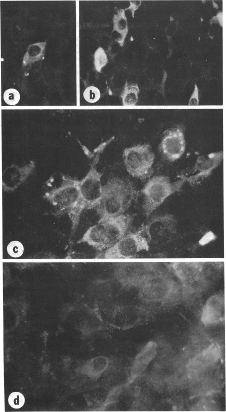
An immunofluorescent cell (IFC) assay technique was developed for the quantification of infectious pancreatic necrosis (IPN) virus of salmonid fishes. Cover slip cultures of rainbow trout gonad (RTG-2) cells were infected with diluted virus preparations. After incubation to permit antigen development, the cells were stained with antiviral fluorescent antibody, and the number of fluorescing (infected) cells was counted. Optimal conditions for the IFC assay procedure are: (i) the use of RTG-2 cells cultured for at least 3 days at 20 C; (ii) 1-h absorption of IPN virus to RTG-2 cells at 20 C or alternatively, 4 h at 4 C; (iii) staining the infected cell cultures at 10 to 12 h postinfection. A linear relationship between the relative concentration of virus in the inoculum and the number of fluorescent cells in the first cycle of infection was observed. The IFC assay method is more sensitive than the plaque method for the assay of IPN virus.
开发了一种免疫荧光细胞(IFC)检测技术,用于定量鲑科鱼类的传染性胰腺坏死(IPN)病毒。用稀释的病毒制剂感染虹鳟性腺(RTG-2)细胞的盖玻片培养物。孵育以促进抗原形成后,用抗病毒荧光抗体对细胞进行染色,并对发荧光(感染)的细胞数量进行计数。IFC检测程序的最佳条件是:(i)使用在20℃下培养至少3天的RTG-2细胞;(ii)在20℃下将IPN病毒吸附到RTG-2细胞上1小时,或者在4℃下吸附4小时;(iii)在感染后10至12小时对感染的细胞培养物进行染色。观察到接种物中病毒的相对浓度与感染第一个周期中荧光细胞数量之间存在线性关系。IFC检测方法在检测IPN病毒方面比噬斑法更灵敏。