• 文献检索
  • 文档翻译
  • 深度研究
  • 学术资讯
  • Suppr Zotero 插件Zotero 插件
  • 邀请有礼
  • 套餐&价格
  • 历史记录
应用&插件
Suppr Zotero 插件Zotero 插件浏览器插件Mac 客户端Windows 客户端微信小程序
定价
高级版会员购买积分包购买API积分包
服务
文献检索文档翻译深度研究API 文档MCP 服务
关于我们
关于 Suppr公司介绍联系我们用户协议隐私条款
关注我们

Suppr 超能文献

核心技术专利:CN118964589B侵权必究
粤ICP备2023148730 号-1Suppr @ 2026

文献检索

告别复杂PubMed语法,用中文像聊天一样搜索,搜遍4000万医学文献。AI智能推荐,让科研检索更轻松。

立即免费搜索

文件翻译

保留排版,准确专业,支持PDF/Word/PPT等文件格式,支持 12+语言互译。

免费翻译文档

深度研究

AI帮你快速写综述,25分钟生成高质量综述,智能提取关键信息,辅助科研写作。

立即免费体验

大肠杆菌B株和B-r株同步生长期间隔膜形成的电子显微镜研究

Electron microscope study of septum formation in Escherichia coli strains B and B-r during synchronous growth.

作者信息

Burdett I D, Murray R G

出版信息

J Bacteriol. 1974 Sep;119(3):1039-56. doi: 10.1128/jb.119.3.1039-1056.1974.

DOI:10.1128/jb.119.3.1039-1056.1974
PMID:4604418
原文链接:https://pmc.ncbi.nlm.nih.gov/articles/PMC245711/
Abstract

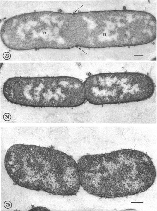
The formation of cell wall septa was monitored in Escherichia coli B and B/r during synchronous growth in glucose media at 37 C by means of electron microscopy. The visible events of septation comprised the following sequence, starting at about 30 min of incubation: (a) bleb formation of the outer membrane; (b) invagination of mucopeptide and cytoplasmic membrane (with associated mesosomes); the outer membrane is excluded from the septum; (c) formation of a cross-wall; (d) ingrowth of the outer membrane during cell separation. The septum is composed of a fold of cytoplasmic membrane plus mucopeptide, and the latter is a double structure, composed of two opposed lamellae separated by an electron-transparent gap. Experiments with chloramphenicol and nalidixic acid suggested that division could occur in the presence of these inhibitors once a round of deoxyribonucleic acid replication is completed. The initial stages of septation, as estimated by the potential of the cells to produce bulges in the presence of ampicillin, may involve the modification of mucopeptide by hydrolases at the end of the C period. Assembly of the septum may occur during the first half of the D period by means of precursors synthesized during the preceding C period.

摘要

通过电子显微镜监测了大肠杆菌B和B/r在37℃的葡萄糖培养基中同步生长期间细胞壁隔膜的形成。隔膜形成的可见事件包括以下顺序,从培养约30分钟开始:(a) 外膜形成泡;(b) 粘肽和细胞质膜(伴有间体)内陷;外膜被排除在隔膜之外;(c) 形成横壁;(d) 细胞分离期间外膜向内生长。隔膜由细胞质膜折叠加上粘肽组成,后者是一种双重结构,由两个相对的薄片组成,中间有一个电子透明间隙。氯霉素和萘啶酸实验表明,一旦一轮脱氧核糖核酸复制完成,在这些抑制剂存在的情况下仍可发生分裂。根据细胞在氨苄青霉素存在下产生凸起的能力估计,隔膜形成的初始阶段可能涉及C期末水解酶对粘肽的修饰。隔膜的组装可能在D期的前半段通过前一个C期合成的前体进行。

https://cdn.ncbi.nlm.nih.gov/pmc/blobs/50a3/245711/7a1c8f16c108/jbacter00339-0414-a.jpg
https://cdn.ncbi.nlm.nih.gov/pmc/blobs/50a3/245711/b1f8e12ed326/jbacter00339-0405-a.jpg
https://cdn.ncbi.nlm.nih.gov/pmc/blobs/50a3/245711/90dff17f0912/jbacter00339-0406-a.jpg
https://cdn.ncbi.nlm.nih.gov/pmc/blobs/50a3/245711/7221e2cf234b/jbacter00339-0408-a.jpg
https://cdn.ncbi.nlm.nih.gov/pmc/blobs/50a3/245711/c1fd1b2e8bb6/jbacter00339-0409-a.jpg
https://cdn.ncbi.nlm.nih.gov/pmc/blobs/50a3/245711/2b761603ad83/jbacter00339-0410-a.jpg
https://cdn.ncbi.nlm.nih.gov/pmc/blobs/50a3/245711/38fce8e50ad3/jbacter00339-0411-a.jpg
https://cdn.ncbi.nlm.nih.gov/pmc/blobs/50a3/245711/7a1c8f16c108/jbacter00339-0414-a.jpg

相似文献

1
Electron microscope study of septum formation in Escherichia coli strains B and B-r during synchronous growth.大肠杆菌B株和B-r株同步生长期间隔膜形成的电子显微镜研究
J Bacteriol. 1974 Sep;119(3):1039-56. doi: 10.1128/jb.119.3.1039-1056.1974.
2
Septum formation in Escherichia coli: characterization of septal structure and the effects of antibiotics on cell division.大肠杆菌中隔膜的形成:隔膜结构的表征以及抗生素对细胞分裂的影响
J Bacteriol. 1974 Jul;119(1):303-24. doi: 10.1128/jb.119.1.303-324.1974.
3
Temperature-sensitive divisionless mutant of Bacillus subtilis defective in the initiation of septation.枯草芽孢杆菌的温度敏感型无分裂突变体,在隔膜形成起始阶段存在缺陷。
J Bacteriol. 1973 Feb;113(2):985-98. doi: 10.1128/jb.113.2.985-998.1973.
4
Cell division in a chain-forming envA mutant of Escherichia coli K12. Fine structure of division sites and effects of EDTA, lysozyme and ampicillin.大肠杆菌K12链形成envA突变体中的细胞分裂。分裂位点的精细结构以及EDTA、溶菌酶和氨苄青霉素的作用。
Acta Pathol Microbiol Scand B Microbiol Immunol. 1971;79(5):651-64. doi: 10.1111/j.1699-0463.1971.tb00093.x.
5
Inhibition of an early event in the cell division cycle of Escherichia coli by FL1060, an amidinopenicillanic acid.脒基青霉烷酸FL1060对大肠杆菌细胞分裂周期早期事件的抑制作用。
J Bacteriol. 1975 Jun;122(3):1283-92. doi: 10.1128/jb.122.3.1283-1292.1975.
6
Coupling between chromosome completion and cell division in Escherichia coli.大肠杆菌中染色体完成与细胞分裂之间的偶联
J Bacteriol. 1973 Sep;115(3):786-95. doi: 10.1128/jb.115.3.786-795.1973.
7
Induction of cell division in a temperature-sensitive division mutant of Escherichia coli by inhibition of protein synthesis.通过抑制蛋白质合成诱导大肠杆菌温度敏感型分裂突变体中的细胞分裂。
J Gen Microbiol. 1977 Apr;99(2):283-90. doi: 10.1099/00221287-99-2-283.
8
Effect of nalidixic acid and hydroxyurea on division ability of Escherichia coli fil+ and lon- strains.萘啶酸和羟基脲对大肠杆菌fil +和lon -菌株分裂能力的影响。
J Bacteriol. 1968 Feb;95(2):520-30. doi: 10.1128/jb.95.2.520-530.1968.
9
Identification of an outer membrane protein of Escherichia coli, with a role in the coordination of deoxyribonucleic acid replication and cell elongation.鉴定大肠杆菌的一种外膜蛋白,其在脱氧核糖核酸复制和细胞伸长的协调中起作用。
J Bacteriol. 1975 Nov;124(2):918-29. doi: 10.1128/jb.124.2.918-929.1975.
10
Evidence for a role of N-acetylmuramyl-L-alanine amidase in septum separation in Escherichia coli.N-乙酰胞壁酰-L-丙氨酸酰胺酶在大肠杆菌隔膜分离中作用的证据。
J Bacteriol. 1976 Nov;128(2):580-6. doi: 10.1128/jb.128.2.580-586.1976.

引用本文的文献

1
Membrane vesicles in class extreme acidophiles: influence on collective behaviors of ''.极端嗜酸菌中的膜泡:对“……集体行为的影响”
Front Microbiol. 2024 Jan 26;14:1331363. doi: 10.3389/fmicb.2023.1331363. eCollection 2023.
2
Emerging role of microbiota derived outer membrane vesicles to preventive, therapeutic and diagnostic proposes.微生物群衍生的外膜囊泡在预防、治疗和诊断方面的新作用。
Infect Agent Cancer. 2023 Jan 19;18(1):3. doi: 10.1186/s13027-023-00480-4.
3
Cell wall synthesis and remodelling dynamics determine division site architecture and cell shape in Escherichia coli.

本文引用的文献

1
Regulation of Cell Division in Escherichia coli: Characterization of Temperature-Sensitive Division Mutants.大肠杆菌中细胞分裂的调控:温度敏感型分裂突变体的特征分析。
J Bacteriol. 1970 Dec;104(3):1052-64. doi: 10.1128/jb.104.3.1052-1064.1970.
2
THE LOCATION OF THE MUCOPEPTIDE IN SECTIONS OF THE CELL WALL OF ESCHERICHIA COLI AND OTHER GRAM-NEGATIVE BACTERIA.黏肽在大肠杆菌及其他革兰氏阴性菌细胞壁切片中的定位。
Can J Microbiol. 1965 Jun;11:547-60. doi: 10.1139/m65-072.
3
TIME-LAPSE PHOTOMICROGRAPHY OF CELL GROWTH AND DIVISION IN ESCHERICHIA COLI.
细胞壁的合成和重塑动态决定了大肠杆菌中分裂位点的结构和细胞形状。
Nat Microbiol. 2022 Oct;7(10):1621-1634. doi: 10.1038/s41564-022-01210-z. Epub 2022 Sep 12.
4
Bacterial extracellular vesicles as bioactive nanocarriers for drug delivery: Advances and perspectives.细菌细胞外囊泡作为药物递送的生物活性纳米载体:进展与展望
Bioact Mater. 2021 Dec 17;14:169-181. doi: 10.1016/j.bioactmat.2021.12.006. eCollection 2022 Aug.
5
Gene Transfer Potential of Outer Membrane Vesicles of Gram-Negative Bacteria.革兰氏阴性菌外膜囊泡的基因转移潜力。
Int J Mol Sci. 2021 Jun 1;22(11):5985. doi: 10.3390/ijms22115985.
6
The role of Zur-regulated lipoprotein A in bacterial morphology, antimicrobial susceptibility, and production of outer membrane vesicles in Acinetobacter baumannii.Zur 调控的脂多糖 A 在鲍曼不动杆菌的细菌形态、抗菌药物敏感性和外膜囊泡产生中的作用。
BMC Microbiol. 2021 Jan 18;21(1):27. doi: 10.1186/s12866-020-02083-0.
7
Eco-evolutionary feedbacks mediated by bacterial membrane vesicles.由细菌膜泡介导的生态进化反馈。
FEMS Microbiol Rev. 2021 Mar 16;45(2). doi: 10.1093/femsre/fuaa047.
8
Fundamental principles in bacterial physiology-history, recent progress, and the future with focus on cell size control: a review.细菌生理学基础——历史、最新进展及未来展望,重点关注细胞大小控制:综述。
Rep Prog Phys. 2018 May;81(5):056601. doi: 10.1088/1361-6633/aaa628. Epub 2018 Jan 9.
9
Real-time detection of antibiotic activity by measuring nanometer-scale bacterial deformation.通过测量纳米级细菌变形实时检测抗生素活性。
J Biomed Opt. 2017 Dec;22(12):1-9. doi: 10.1117/1.JBO.22.12.126002.
10
Outer Membrane Vesicles (OMVs) of Gram-negative Bacteria: A Perspective Update.革兰氏阴性菌的外膜囊泡:视角更新
Front Microbiol. 2017 Jun 9;8:1053. doi: 10.3389/fmicb.2017.01053. eCollection 2017.
大肠杆菌细胞生长与分裂的延时显微摄影
J Bacteriol. 1965 Jan;89(1):212-6. doi: 10.1128/jb.89.1.212-216.1965.
4
AN IMPROVED METHOD FOR THE SELECTION OF BACTERIAL CELLS AT DIVISION.一种用于选择处于分裂期细菌细胞的改进方法。
Biochim Biophys Acta. 1964 Mar 16;82:608-10. doi: 10.1016/0304-4165(64)90453-2.
5
The molecular synchrony and sequential replication of DNA in Escherichia coli.大肠杆菌中DNA的分子同步性和序列复制
Proc Natl Acad Sci U S A. 1963 Apr;49(4):551-9. doi: 10.1073/pnas.49.4.551.
6
[Electron microscopic study on plasmas containing desoxyribonucleic acid. I. Nucleoids of actively growing bacteria].[含脱氧核糖核酸血浆的电子显微镜研究。I. 活跃生长细菌的拟核]
Z Naturforsch B. 1958 Sep;13B(9):597-605.
7
The fine structure of Thioploca ingrica and a comparison with Beggiatoa.英格里卡硫丝菌的精细结构及其与贝氏硫菌的比较。
Can J Microbiol. 1965 Aug;11(4):645-55. doi: 10.1139/m65-087.
8
The cell wall and cell division of gram-negative bacteria.革兰氏阴性菌的细胞壁与细胞分裂
Can J Microbiol. 1966 Apr;12(2):263-70. doi: 10.1139/m66-036.
9
Procaryotic cell division with respect to wall and membranes.原核细胞在细胞壁和细胞膜方面的分裂
CRC Crit Rev Microbiol. 1971 May;1(1):29-72. doi: 10.3109/10408417109104477.
10
Lipid synthesis in relation to the cell cycle of Bacillus megaterium KM and Escherichia coli.巨大芽孢杆菌KM和大肠杆菌的脂质合成与细胞周期的关系
Biochem J. 1969 Dec;115(4):697-701. doi: 10.1042/bj1150697.