Nelson J B, Lyons M J
J Bacteriol. 1965 Dec;90(6):1750-63. doi: 10.1128/jb.90.6.1750-1763.1965.
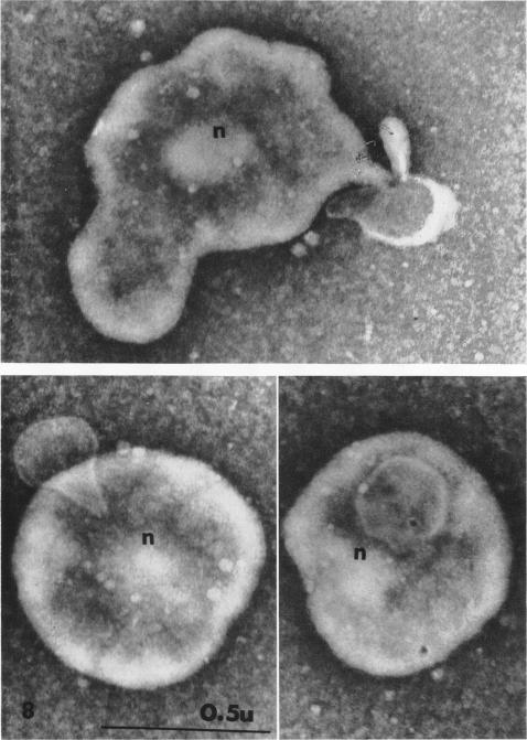
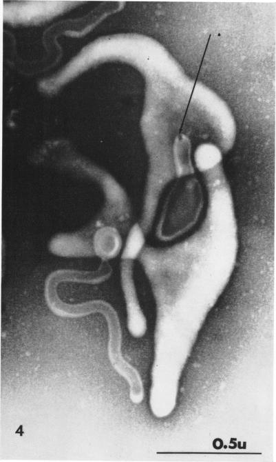
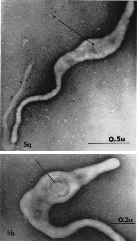
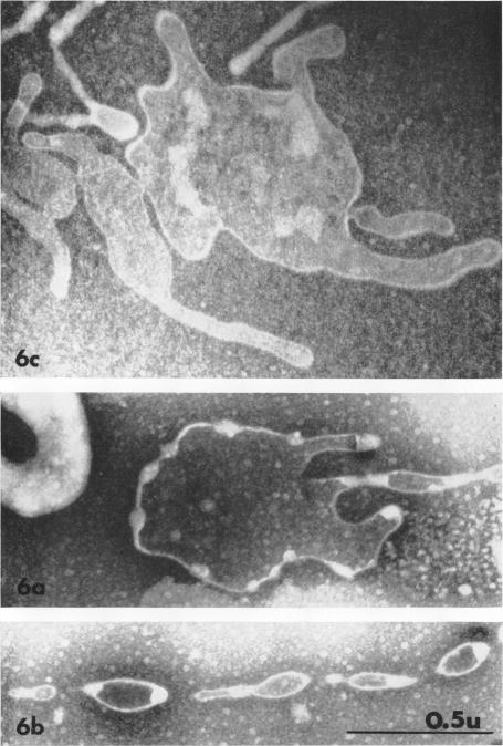
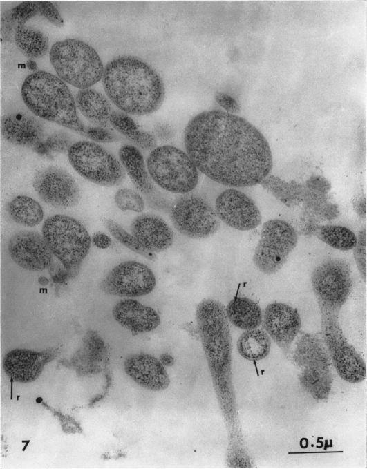
Nelson, John B. (The Rockefeller University, New York, N.Y.), and Michael J. Lyons. Phase-contrast and electron microscopy of murine strains of Mycoplasma. J. Bacteriol. 90:1750-1763. 1965.-Two strains of Mycoplasma pulmonis (associated with infectious catarrh) on examination in fluid culture (20% horse serum-bouillon) by phase microscopy were highly pleomorphic, with many bacilliform elements and fewer coccoid ones. Motility, characterized by gliding of rods and spinning of spherical forms, was observed through the 9th subculture of one strain and the 15th of the second. Motile elements were not seen in later transfers and pleomorphism was reduced. One strain of M. neurolyticum (associated with conjunctivitis and encephalitis) was much less pleomorphic and showed neither bacilliform elements nor motility at any time. When examined by negative-contrast electron microscopy, organisms of this strain were found to have an average diameter of 0.7 mu and to possess a concentrated peripheral layer of cytoplasm and a central mass which may represent the cells' nuclear equivalent. The latter feature was not prominent in spherical forms of M. pulmonis. These cells, when observed after 48 hr of culture, showed evidence of the generation of new progeny cells in their central area. The filamentous or bacilliform cells of M. pulmonis were frequently serpentine in appearance, 2.0 to 3.0 mu in length and 80 to 250 mmu in width. They appeared to generate new cells from terminal buds from which outpouchings initially developed. Older cells, in the stationary phase, showed evidence of undergoing multipolar germination. Microtubules, about 60 mmu wide, were found in association with most filamentous cells from 48-hr cultures; fragments of membrane, studded with closely packed ribosomelike particles, were also found. There was no evidence of flagella or any specialized structure that could account for the observed motility of the organisms.
尼尔森,约翰·B.(纽约洛克菲勒大学)和迈克尔·J.莱昂斯。鼠源支原体菌株的相差显微镜和电子显微镜观察。《细菌学杂志》90:1750 - 1763。1965年。——两株肺支原体(与传染性卡他有关)在液体培养基(20%马血清肉汤)中通过相差显微镜检查时高度多形性,有许多杆状形态,较少球状形态。在一株的第9代传代培养物和另一株的第15代传代培养物中观察到运动性,其特征为杆状的滑动和球状的旋转。在后续传代中未观察到运动性成分,多形性也有所降低。一株溶神经支原体(与结膜炎和脑炎有关)的多形性要小得多,在任何时候都未显示杆状形态或运动性。通过负染色电子显微镜检查发现,该菌株的生物体平均直径为0.7微米,具有一层浓缩的外周细胞质层和一个中央团块,可能相当于细胞的核。后一特征在肺支原体的球状形态中不明显。这些细胞在培养48小时后观察时,在其中心区域显示出新子代细胞产生的迹象。肺支原体的丝状或杆状细胞外观常呈蜿蜒状,长2.0至3.0微米,宽80至250毫微米。它们似乎从最初出现外突的末端芽产生新细胞。处于稳定期的老龄细胞显示出多极萌发的迹象。在48小时培养物的大多数丝状细胞中发现了约60毫微米宽的微管;还发现了布满紧密排列的核糖体样颗粒的膜碎片。没有证据表明存在鞭毛或任何能解释所观察到的生物体运动性的特殊结构。