• 文献检索
  • 文档翻译
  • 深度研究
  • 学术资讯
  • Suppr Zotero 插件Zotero 插件
  • 邀请有礼
  • 套餐&价格
  • 历史记录
应用&插件
Suppr Zotero 插件Zotero 插件浏览器插件Mac 客户端Windows 客户端微信小程序
定价
高级版会员购买积分包购买API积分包
服务
文献检索文档翻译深度研究API 文档MCP 服务
关于我们
关于 Suppr公司介绍联系我们用户协议隐私条款
关注我们

Suppr 超能文献

核心技术专利:CN118964589B侵权必究
粤ICP备2023148730 号-1Suppr @ 2026

文献检索

告别复杂PubMed语法,用中文像聊天一样搜索,搜遍4000万医学文献。AI智能推荐,让科研检索更轻松。

立即免费搜索

文件翻译

保留排版,准确专业,支持PDF/Word/PPT等文件格式,支持 12+语言互译。

免费翻译文档

深度研究

AI帮你快速写综述,25分钟生成高质量综述,智能提取关键信息,辅助科研写作。

立即免费体验

腐败厌氧菌3679h在孢子形成过程中的超微结构

Ultrastructure of putrefactive anaerobe 3679h during sporulation.

作者信息

Santo L M, Hohl H R, Frank H A

出版信息

J Bacteriol. 1969 Sep;99(3):824-33. doi: 10.1128/jb.99.3.824-833.1969.

DOI:10.1128/jb.99.3.824-833.1969
PMID:5370279
原文链接:https://pmc.ncbi.nlm.nih.gov/articles/PMC250100/
Abstract

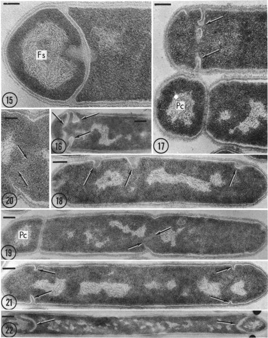
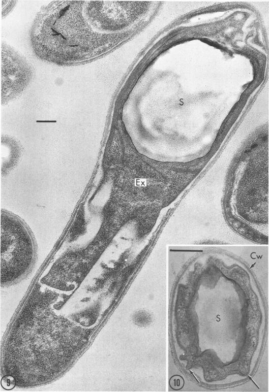
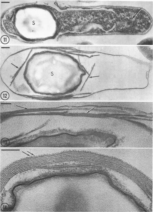
Sporulation of putrefactive anaerobe 3679h was studied by observation of ultra-thin sections in the electron microscope. The customary stages were observed during forespore formation, spore maturation, and liberation of the free spore. The exosporium was partially laminated and devoid of the hairlike surface projections observed on spores of some species. Atypical membrane configurations occurred in some cells, and, occasionally, cells with bipolar forespores were found.

摘要

通过电子显微镜观察超薄切片,对腐败厌氧菌3679h的芽孢形成进行了研究。在前芽孢形成、芽孢成熟和游离芽孢释放过程中观察到了常见阶段。芽孢衣部分呈层状,且没有在某些物种的芽孢上观察到的毛发状表面突起。在一些细胞中出现了非典型的膜结构,偶尔还发现了带有双极前芽孢的细胞。

https://cdn.ncbi.nlm.nih.gov/pmc/blobs/8933/250100/07e43a1faeaf/jbacter00389-0237-a.jpg
https://cdn.ncbi.nlm.nih.gov/pmc/blobs/8933/250100/374457f1a842/jbacter00389-0232-a.jpg
https://cdn.ncbi.nlm.nih.gov/pmc/blobs/8933/250100/672bad430415/jbacter00389-0233-a.jpg
https://cdn.ncbi.nlm.nih.gov/pmc/blobs/8933/250100/04d8429cd64c/jbacter00389-0234-a.jpg
https://cdn.ncbi.nlm.nih.gov/pmc/blobs/8933/250100/b3152bb7c3cc/jbacter00389-0236-a.jpg
https://cdn.ncbi.nlm.nih.gov/pmc/blobs/8933/250100/07e43a1faeaf/jbacter00389-0237-a.jpg

相似文献

1
Ultrastructure of putrefactive anaerobe 3679h during sporulation.腐败厌氧菌3679h在孢子形成过程中的超微结构
J Bacteriol. 1969 Sep;99(3):824-33. doi: 10.1128/jb.99.3.824-833.1969.
2
Ultrastructure of the exosporium and underlying inclusions in spores of Bacillus megaterium strains.巨大芽孢杆菌菌株孢子中芽孢外壁及内部包含物的超微结构
J Bacteriol. 1972 Mar;109(3):1198-209. doi: 10.1128/jb.109.3.1198-1209.1972.
3
Ultrastructural changes associated with germination and outgrowth of an appendage-bearing clostridial spore.与带有附属物的梭菌芽孢萌发和生长相关的超微结构变化。
J Bacteriol. 1970 Mar;101(3):1038-45. doi: 10.1128/jb.101.3.1038-1045.1970.
4
Exosporium and spore coat formation in Bacillus cereus T.蜡样芽孢杆菌T中芽孢外壁和芽孢衣的形成
J Bacteriol. 1973 Sep;115(3):1179-90. doi: 10.1128/jb.115.3.1179-1190.1973.
5
ULTRASTRUCTURE OF THE EXOSPORIUM ENVELOPING SPORES OF BACILLUS CEREUS.蜡样芽孢杆菌芽孢外孢膜的超微结构
J Bacteriol. 1964 Dec;88(6):1774-89. doi: 10.1128/jb.88.6.1774-1789.1964.
6
Sequence of events during germination of putrefactive anaerobe 3679 spores.腐败厌氧菌3679孢子萌发过程中的事件顺序。
J Bacteriol. 1967 Sep;94(3):506-11. doi: 10.1128/jb.94.3.506-511.1967.
7
Formation and structure of the spore of Bacillus coagulans.凝结芽孢杆菌芽孢的形成与结构。
J Cell Biol. 1962 Jul;14(1):111-23. doi: 10.1083/jcb.14.1.111.
8
Sporulation in Bacillus subtilis. Morphological changes.枯草芽孢杆菌中的孢子形成。形态变化。
Biochem J. 1968 Oct;109(5):819-24. doi: 10.1042/bj1090819.
9
Appendage development in Clostridium bifermentans.双酶梭菌的附属器发育
J Bacteriol. 1971 Apr;106(1):269-75. doi: 10.1128/jb.106.1.269-275.1971.
10
Fine structure of mesosomal involvement during Bacillus macerans sporulation.浸麻芽孢杆菌芽孢形成过程中中间体参与的精细结构
J Bacteriol. 1975 Jan;121(1):363-72. doi: 10.1128/jb.121.1.363-372.1975.

引用本文的文献

1
Ultrastructural changes associated with germination and outgrowth of an appendage-bearing clostridial spore.与带有附属物的梭菌芽孢萌发和生长相关的超微结构变化。
J Bacteriol. 1970 Mar;101(3):1038-45. doi: 10.1128/jb.101.3.1038-1045.1970.
2
Exosporium formation in sporulating cells of Clostridium botulinum 78A.肉毒梭菌78A芽孢形成细胞中的芽孢外壁形成
J Bacteriol. 1972 Oct;112(1):618-21. doi: 10.1128/jb.112.1.618-621.1972.
3
Interference-contrast and phase-contrast microscopy of sporulation in clostridium thermosaccharolyticum grown under strict anaerobiosis.

本文引用的文献

1
MORPHOLOGY OF SPORE DEVELOPMENT IN CLOSTRIDIUM PECTINOVORUM.果胶分解梭菌孢子发育的形态学
J Bacteriol. 1962 Jul;84(1):104-14. doi: 10.1128/jb.84.1.104-114.1962.
2
[MORPHOLOGIC STUDY OF THE SPORULATION OF BACILLUS SUBTILIS].[枯草芽孢杆菌芽孢形成的形态学研究]
Ann Inst Pasteur (Paris). 1965 Jan;108:40-60.
3
METHOD FOR OBTAINING CLEANED PUTREFACTIVE ANAEROBE 3679 SPORES.获取经清洁处理的腐败厌氧菌3679孢子的方法。
在严格厌氧条件下生长的嗜热解糖梭菌孢子形成的干涉对比显微镜和相差显微镜观察
J Bacteriol. 1972 Sep;111(3):674-81. doi: 10.1128/jb.111.3.674-681.1972.
4
Exosporium and spore coat formation in Bacillus cereus T.蜡样芽孢杆菌T中芽孢外壁和芽孢衣的形成
J Bacteriol. 1973 Sep;115(3):1179-90. doi: 10.1128/jb.115.3.1179-1190.1973.
5
Ultrastructure of the exosporium and underlying inclusions in spores of Bacillus megaterium strains.巨大芽孢杆菌菌株孢子中芽孢外壁及内部包含物的超微结构
J Bacteriol. 1972 Mar;109(3):1198-209. doi: 10.1128/jb.109.3.1198-1209.1972.
6
Appendage development in Clostridium bifermentans.双酶梭菌的附属器发育
J Bacteriol. 1971 Apr;106(1):269-75. doi: 10.1128/jb.106.1.269-275.1971.
7
Sheathed cells in cultures of Clostridium sporogenes.产孢梭菌培养物中的鞘细胞。
J Bacteriol. 1970 Sep;103(3):814-25. doi: 10.1128/jb.103.3.814-825.1970.
J Bacteriol. 1965 Mar;89(3):929-30. doi: 10.1128/jb.89.3.929-930.1965.
4
ULTRASTRUCTURE OF THE EXOSPORIUM ENVELOPING SPORES OF BACILLUS CEREUS.蜡样芽孢杆菌芽孢外孢膜的超微结构
J Bacteriol. 1964 Dec;88(6):1774-89. doi: 10.1128/jb.88.6.1774-1789.1964.
5
CHARACTERISTICS OF AN ABORTIVELY DISPORIC VARIANT OF BACILLUS CEREUS.蜡样芽孢杆菌流产性双芽孢变种的特征
J Bacteriol. 1964 Jul;88(1):242-54. doi: 10.1128/jb.88.1.242-254.1964.
6
ELECTRON MICROSCOPY OF SPORE FORMATION AND GERMINATION IN BACILLUS SUBTILIS.
Jpn J Microbiol. 1963 Jun;7:23-41. doi: 10.1111/j.1348-0421.1963.tb00926.x.
7
The use of lead citrate at high pH as an electron-opaque stain in electron microscopy.在电子显微镜检查中,将高pH值的柠檬酸铅用作电子不透明染色剂。
J Cell Biol. 1963 Apr;17(1):208-12. doi: 10.1083/jcb.17.1.208.
8
Electron microscope studies on ultrathin sections of spores of the Clostridium group, with special reference to the sporulation and germination process.关于梭菌属孢子超薄切片的电子显微镜研究,特别涉及孢子形成和萌发过程。
J Bacteriol. 1960 Jul;80(1):37-46. doi: 10.1128/jb.80.1.37-46.1960.
9
Electron microscopic studies on ultrathin sections of spores of Clostridium tetani and Clostridium histolyticum, with special reference to sporulation and spore germination process.破伤风梭菌和溶组织梭菌孢子超薄切片的电子显微镜研究,特别涉及孢子形成和孢子萌发过程。
Jpn J Microbiol. 1960 Apr;4:137-55. doi: 10.1111/j.1348-0421.1960.tb00162.x.
10
Participation of the cytoplasmic membrane in the growth and spore fromation of bacilli.细胞质膜在杆菌生长和孢子形成中的作用。
J Biophys Biochem Cytol. 1960 Oct;8(2):507-28. doi: 10.1083/jcb.8.2.507.