• 文献检索
  • 文档翻译
  • 深度研究
  • 学术资讯
  • Suppr Zotero 插件Zotero 插件
  • 邀请有礼
  • 套餐&价格
  • 历史记录
应用&插件
Suppr Zotero 插件Zotero 插件浏览器插件Mac 客户端Windows 客户端微信小程序
定价
高级版会员购买积分包购买API积分包
服务
文献检索文档翻译深度研究API 文档MCP 服务
关于我们
关于 Suppr公司介绍联系我们用户协议隐私条款
关注我们

Suppr 超能文献

核心技术专利:CN118964589B侵权必究
粤ICP备2023148730 号-1Suppr @ 2026

文献检索

告别复杂PubMed语法,用中文像聊天一样搜索,搜遍4000万医学文献。AI智能推荐,让科研检索更轻松。

立即免费搜索

文件翻译

保留排版,准确专业,支持PDF/Word/PPT等文件格式,支持 12+语言互译。

免费翻译文档

深度研究

AI帮你快速写综述,25分钟生成高质量综述,智能提取关键信息,辅助科研写作。

立即免费体验

运动外硫红螺菌(Ectothiorhodospira mobilis Pelsh)的精细结构

Fine structure of Ectothiorhodospira mobilis Pelsh.

作者信息

Remsen C C, Watson S W, Waterbury J B, Trüper H G

出版信息

J Bacteriol. 1968 Jun;95(6):2374-92. doi: 10.1128/jb.95.6.2374-2392.1968.

DOI:10.1128/jb.95.6.2374-2392.1968
PMID:5669908
原文链接:https://pmc.ncbi.nlm.nih.gov/articles/PMC315173/
Abstract

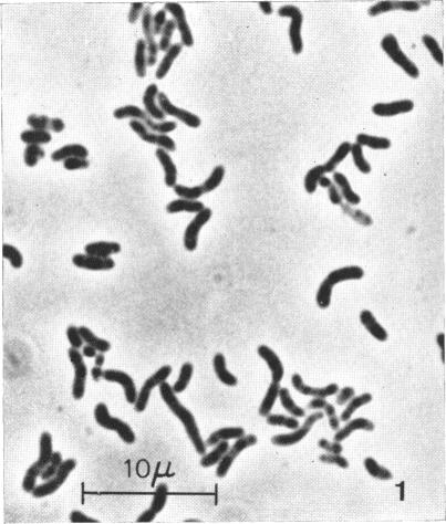
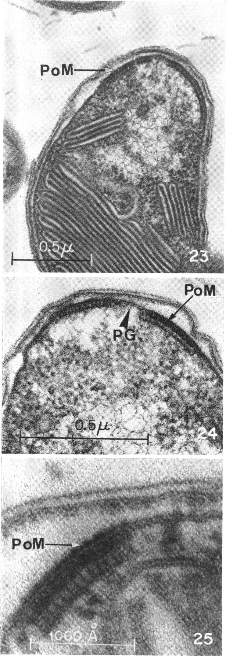
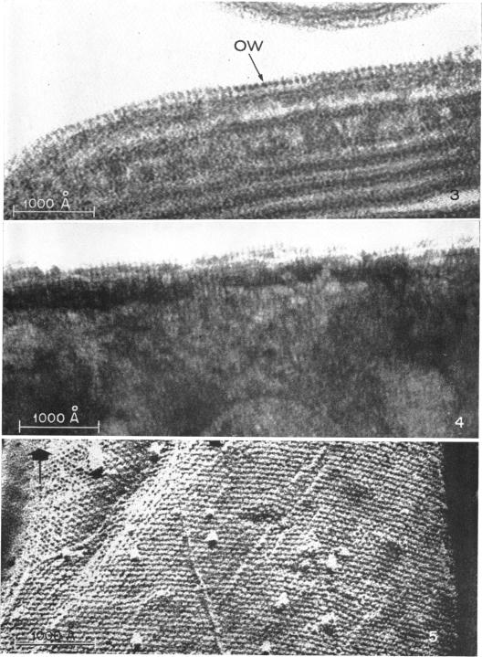
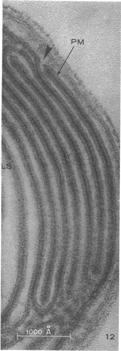
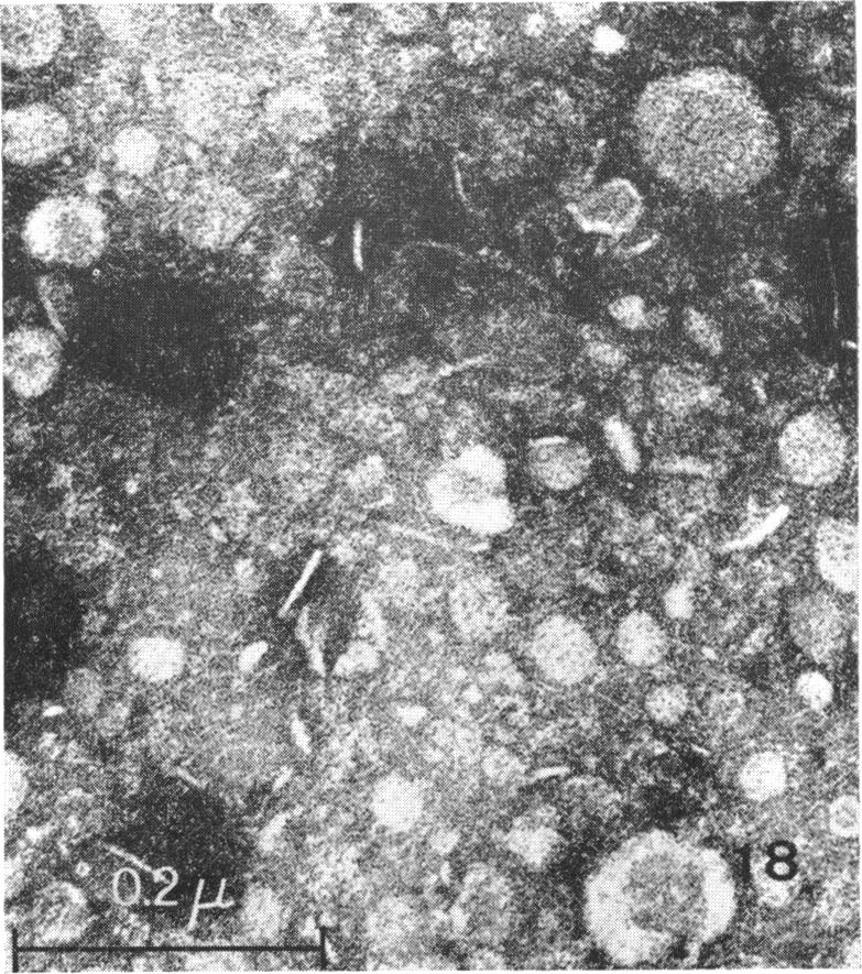
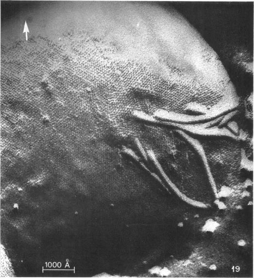
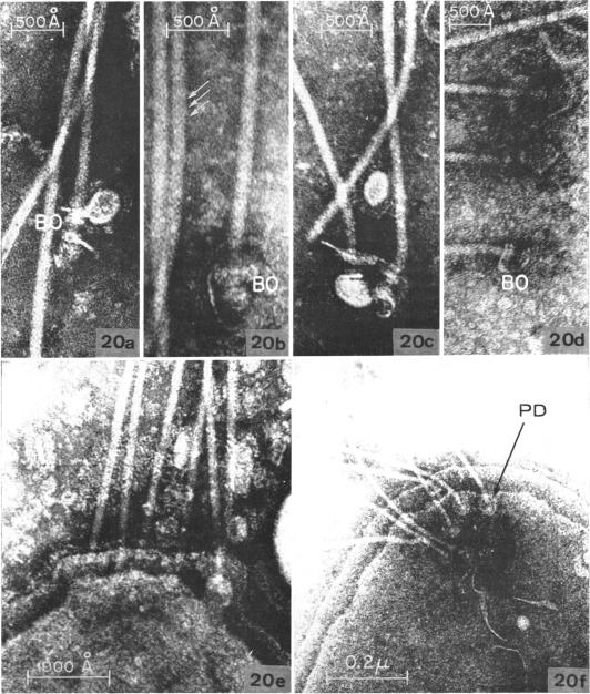
The cell wall structure, arrangement of photosynthetic membranes, and the attachment of flagella of Ectothiorhodospira mobilis strain 8112 were examined by using freeze-etching and conventional electron microscopic techniques. The outer coat of the multilayered cell wall is comprised of 50 A repeating subunits, arranged in a regular array. The photosynthetic membranes, which originate from and are attached to the plasma membrane, are arranged in a more complex pattern than previously seen in other bacteria. The tuft of flagella in E. mobilis is inserted into a polar organelle. The relationship of this organelle to the polar membrane and the mechanism of attachment of the flagella to the polar organelle is discussed.

摘要

运用冷冻蚀刻和传统电子显微镜技术,对运动外硫红螺菌8112菌株的细胞壁结构、光合膜排列以及鞭毛附着情况进行了研究。多层细胞壁的外层由50埃的重复亚基组成,呈规则排列。起源于质膜并附着于质膜的光合膜,其排列方式比之前在其他细菌中所见的更为复杂。运动外硫红螺菌的鞭毛束插入到一个极性细胞器中。本文讨论了该细胞器与极性膜的关系以及鞭毛附着于极性细胞器的机制。

https://cdn.ncbi.nlm.nih.gov/pmc/blobs/9d5f/315173/8188be425975/jbacter00588-0441-a.jpg
https://cdn.ncbi.nlm.nih.gov/pmc/blobs/9d5f/315173/ba990c155d6b/jbacter00588-0427-a.jpg
https://cdn.ncbi.nlm.nih.gov/pmc/blobs/9d5f/315173/5582009f76ae/jbacter00588-0428-a.jpg
https://cdn.ncbi.nlm.nih.gov/pmc/blobs/9d5f/315173/d5ed1cd32116/jbacter00588-0429-a.jpg
https://cdn.ncbi.nlm.nih.gov/pmc/blobs/9d5f/315173/a2113c18852e/jbacter00588-0430-a.jpg
https://cdn.ncbi.nlm.nih.gov/pmc/blobs/9d5f/315173/45d21f3a6815/jbacter00588-0431-a.jpg
https://cdn.ncbi.nlm.nih.gov/pmc/blobs/9d5f/315173/42f61120257c/jbacter00588-0432-a.jpg
https://cdn.ncbi.nlm.nih.gov/pmc/blobs/9d5f/315173/8c3bcd6a2b5c/jbacter00588-0433-a.jpg
https://cdn.ncbi.nlm.nih.gov/pmc/blobs/9d5f/315173/1434e2a24870/jbacter00588-0434-a.jpg
https://cdn.ncbi.nlm.nih.gov/pmc/blobs/9d5f/315173/64895fbfaa1d/jbacter00588-0435-a.jpg
https://cdn.ncbi.nlm.nih.gov/pmc/blobs/9d5f/315173/3320be18420b/jbacter00588-0436-a.jpg
https://cdn.ncbi.nlm.nih.gov/pmc/blobs/9d5f/315173/54f183bd9b05/jbacter00588-0437-a.jpg
https://cdn.ncbi.nlm.nih.gov/pmc/blobs/9d5f/315173/0d1964f305d5/jbacter00588-0438-a.jpg
https://cdn.ncbi.nlm.nih.gov/pmc/blobs/9d5f/315173/1584756e5ed9/jbacter00588-0439-a.jpg
https://cdn.ncbi.nlm.nih.gov/pmc/blobs/9d5f/315173/d1c49024c625/jbacter00588-0440-a.jpg
https://cdn.ncbi.nlm.nih.gov/pmc/blobs/9d5f/315173/8188be425975/jbacter00588-0441-a.jpg

相似文献

1
Fine structure of Ectothiorhodospira mobilis Pelsh.运动外硫红螺菌(Ectothiorhodospira mobilis Pelsh)的精细结构
J Bacteriol. 1968 Jun;95(6):2374-92. doi: 10.1128/jb.95.6.2374-2392.1968.
2
Ectothiorhodospira mobilis Pelsh, a photosynthetic sulfur bacterium depositing sulfur outside the cells.运动外硫红螺菌(Ectothiorhodospira mobilis Pelsh),一种在细胞外沉积硫的光合硫细菌。
J Bacteriol. 1968 May;95(5):1910-20. doi: 10.1128/jb.95.5.1910-1920.1968.
3
Fine structure of Ectothiorhodospira mobilis strain 8113 thylakoids: chemical fixation and freeze-etching studies.运动外硫红螺菌8113菌株类囊体的精细结构:化学固定和冷冻蚀刻研究
Arch Mikrobiol. 1968;62(2):111-28. doi: 10.1007/BF00410398.
4
Freeze-etching of bacteria.
Int Rev Cytol. 1972;33:253-96. doi: 10.1016/s0074-7696(08)61452-7.
5
Relationship between cell wall, cytoplasmic membrane, and bacterial motility.细胞壁、细胞质膜与细菌运动性之间的关系。
J Bacteriol. 1969 Oct;100(1):512-21. doi: 10.1128/jb.100.1.512-521.1969.
6
THE FINE STRUCTURE OF STALKED BACTERIA BELONGING TO THE FAMILY CAULOBACTERACEAE.柄杆菌科柄细菌的精细结构
J Cell Biol. 1964 Dec;23(3):587-607. doi: 10.1083/jcb.23.3.587.
7
[The electron microscopic fine structure of the cell wall, cytoplasmic membrane and flagella of Bacillus stearothermophilus demonstrated by means of freeze etching].嗜热脂肪芽孢杆菌细胞壁、细胞质膜和鞭毛的冷冻蚀刻电子显微镜精细结构
Mikroskopie. 1968 May;22(9):233-42.
8
Freeze-etching of Azotobacter vinelandii: examination of wall, exine, and vesicles.棕色固氮菌的冷冻蚀刻:细胞壁、外壁和囊泡的检查
J Bacteriol. 1972 Mar;109(3):1191-7. doi: 10.1128/jb.109.3.1191-1197.1972.
9
Structure and assembly of bacterial surface layers composed of regular arrays of subunits.由亚基规则排列组成的细菌表面层的结构与组装。
Philos Trans R Soc Lond B Biol Sci. 1974 Jul 25;268(891):147-53. doi: 10.1098/rstb.1974.0022.
10
THE FINE STRUCTURE AND MODE OF ATTACHMENT OF THE SHEATHED FLAGELLUM OF VIBRIO METCHNIKOVII.梅契尼科夫弧菌鞘鞭毛的精细结构及附着方式
J Cell Biol. 1963 Aug;18(2):327-36. doi: 10.1083/jcb.18.2.327.

引用本文的文献

1
A Cristae-Like Microcompartment in .线粒体内嵴样微区结构
mBio. 2022 Dec 20;13(6):e0161322. doi: 10.1128/mbio.01613-22. Epub 2022 Nov 2.
2
Isolation and Characterization of Cell Envelope Fragments Comprising Archaeal S-Layer Proteins.包含古菌S层蛋白的细胞包膜片段的分离与鉴定
Nanomaterials (Basel). 2022 Jul 21;12(14):2502. doi: 10.3390/nano12142502.
3
S-layers: principles and applications.S层:原理与应用

本文引用的文献

1
Photosynthetic Apparatus in the Green Bacterium Chloropseudomonas ethylicum.绿色细菌嗜乙基绿假单胞菌中的光合装置。
J Bacteriol. 1966 Jan;91(1):311-23. doi: 10.1128/jb.91.1.311-323.1966.
2
ULTRASTRUCTURE OF THE CELL WALL OF ESCHERICHIA COLI.大肠杆菌细胞壁的超微结构
J Ultrastruct Res. 1965 Apr;12:247-62. doi: 10.1016/s0022-5320(65)80098-3.
3
STRUCTURE OF NITROSOCYSTIS OCEANUS AND COMPARISON WITH NITROSOMONAS AND NITROBACTER.海洋亚硝化囊菌的结构及其与亚硝化单胞菌和硝化杆菌的比较。
FEMS Microbiol Rev. 2014 Sep;38(5):823-64. doi: 10.1111/1574-6976.12063. Epub 2014 Feb 24.
4
Keys to eukaryality: planctomycetes and ancestral evolution of cellular complexity.真核生物的关键:浮霉菌门与细胞复杂性的祖先进化
Front Microbiol. 2012 May 4;3:167. doi: 10.3389/fmicb.2012.00167. eCollection 2012.
5
Microflora of soil as viewed by transmission electron microscopy.通过透射电子显微镜观察土壤微生物区系。
Appl Microbiol. 1972 Mar;23(3):637-48. doi: 10.1128/am.23.3.637-648.1972.
6
Association of a new type of gliding, filamentous, purple phototrophic bacterium inside bundles of Microcoleus chthonoplastes in hypersaline cyanobacterial mats.一种新型滑行丝状紫色光合细菌与超盐度蓝细菌垫中鞘丝藻束内的细菌的关联。
Arch Microbiol. 1987;147:213-20. doi: 10.1007/BF00463477.
7
Changes in the evolution of the antigenic profiles and morphology during coccoid conversion of Helicobacter pylori.幽门螺杆菌球状体转化过程中抗原谱和形态演变的变化。
Korean J Intern Med. 1999 Jan;14(1):9-14. doi: 10.3904/kjim.1999.14.1.9.
8
Changes in Helicobacter pylori ultrastructure and antigens during conversion from the bacillary to the coccoid form.幽门螺杆菌从杆菌形态转变为球菌形态过程中超微结构和抗原的变化。
Infect Immun. 1996 Jun;64(6):2331-5. doi: 10.1128/iai.64.6.2331-2335.1996.
9
Bioenergetics: the evolution of molecular mechanisms and the development of bioenergetic concepts.生物能量学:分子机制的演变与生物能量学概念的发展
Antonie Van Leeuwenhoek. 1994;65(4):271-84. doi: 10.1007/BF00872213.
10
Ultrastructure of a magnetotactic spirillum.趋磁螺旋菌的超微结构。
J Bacteriol. 1980 Mar;141(3):1399-408. doi: 10.1128/jb.141.3.1399-1408.1980.
J Bacteriol. 1965 Jun;89(6):1594-609. doi: 10.1128/jb.89.6.1594-1609.1965.
4
OBSERVATIONS ON THE STRUCTURE OF RHODOSPIRILLUM RUBUM.关于红螺菌结构的观察
J Cell Biol. 1965 May;25(2):279-91. doi: 10.1083/jcb.25.2.279.
5
ENERGY SUPPLY FOR THE CHEMOAUTOTROPH FERROBACILLUS FERROOXIDANS.化学自养型氧化亚铁硫杆菌的能量供应
J Bacteriol. 1965 Mar;89(3):825-34. doi: 10.1128/jb.89.3.825-834.1965.
6
BIOLOGY OF BUDDING BACTERIA. 3. FINE STRUCTURE OF RHODOMICROBIUM AND HYPHOMICROBIUM SPP.出芽细菌的生物学。3. 红微菌属和生丝微菌属菌种的精细结构
J Bacteriol. 1965 Feb;89(2):503-12. doi: 10.1128/jb.89.2.503-512.1965.
7
A NEW TYPE OF BACTERIAL CELL WALL STRUCTURE REVEALED BY REPLICA TECHNIQUE.复型技术揭示的新型细菌细胞壁结构
Antonie Van Leeuwenhoek. 1964;30:239-48. doi: 10.1007/BF02046729.
8
THE FINE STRUCTURE OF GREEN BACTERIA.绿色细菌的精细结构
J Cell Biol. 1964 Jul;22(1):207-25. doi: 10.1083/jcb.22.1.207.
9
MORPHOLOGICAL AND CHEMICAL PROPERTIES OF CELL ENVELOPES OF THE EXTREME HALOPHILE, HALOBACTERIUM CUTIRUBRUM.极端嗜盐菌——红皮盐杆菌细胞包膜的形态学与化学特性
Can J Microbiol. 1964 Jun;10:483-97. doi: 10.1139/m64-058.
10
The fine structure of Rhodospirillum rubrum.深红红螺菌的精细结构。
J Cell Biol. 1963 Feb;16(2):401-19. doi: 10.1083/jcb.16.2.401.