• 文献检索
  • 文档翻译
  • 深度研究
  • 学术资讯
  • Suppr Zotero 插件Zotero 插件
  • 邀请有礼
  • 套餐&价格
  • 历史记录
应用&插件
Suppr Zotero 插件Zotero 插件浏览器插件Mac 客户端Windows 客户端微信小程序
定价
高级版会员购买积分包购买API积分包
服务
文献检索文档翻译深度研究API 文档MCP 服务
关于我们
关于 Suppr公司介绍联系我们用户协议隐私条款
关注我们

Suppr 超能文献

核心技术专利:CN118964589B侵权必究
粤ICP备2023148730 号-1Suppr @ 2026

文献检索

告别复杂PubMed语法,用中文像聊天一样搜索,搜遍4000万医学文献。AI智能推荐,让科研检索更轻松。

立即免费搜索

文件翻译

保留排版,准确专业,支持PDF/Word/PPT等文件格式,支持 12+语言互译。

免费翻译文档

深度研究

AI帮你快速写综述,25分钟生成高质量综述,智能提取关键信息,辅助科研写作。

立即免费体验

去神经支配的鞘细胞在神经损伤后分泌一种新蛋白质。

Denervated sheath cells secrete a new protein after nerve injury.

作者信息

Skene J H, Shooter E M

出版信息

Proc Natl Acad Sci U S A. 1983 Jul;80(13):4169-73. doi: 10.1073/pnas.80.13.4169.

DOI:10.1073/pnas.80.13.4169
PMID:6575401
原文链接:https://pmc.ncbi.nlm.nih.gov/articles/PMC394222/
Abstract

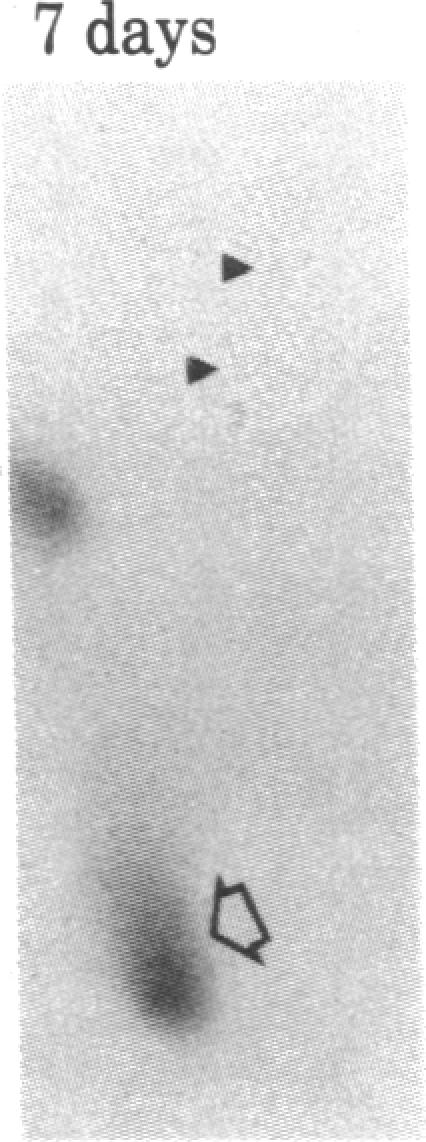
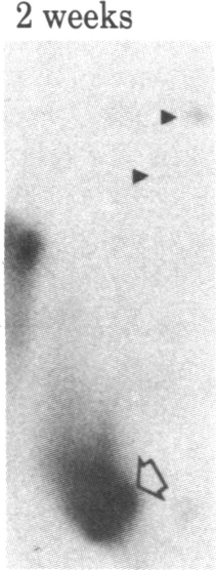
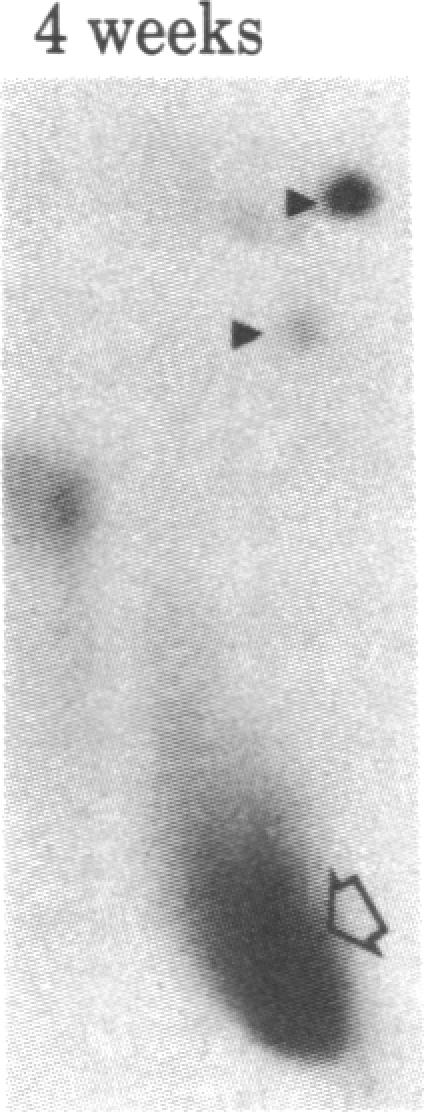
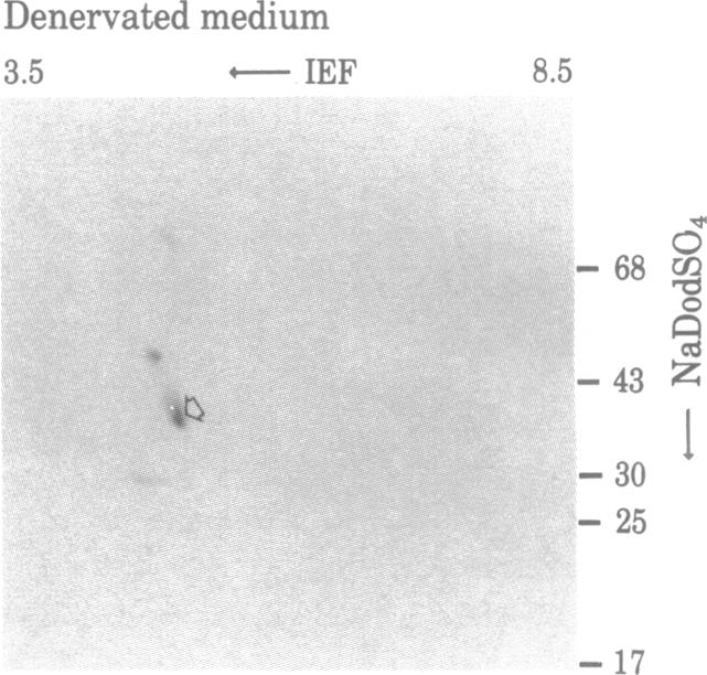
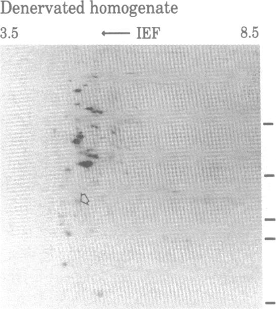
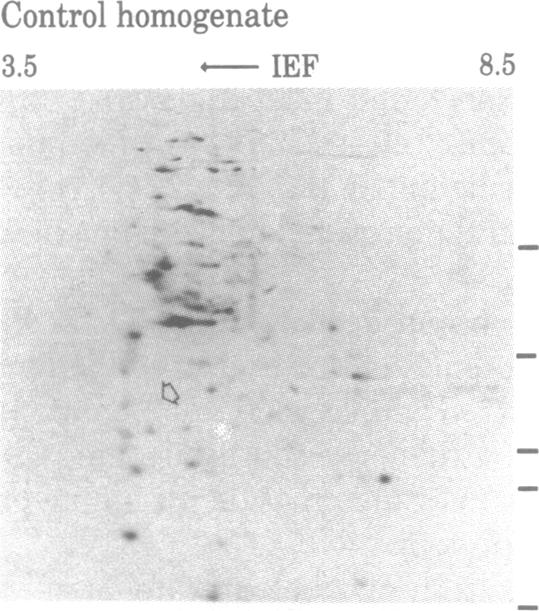
When rat sciatic nerves are crushed, Schwann cells or other supporting cells distal to the injury site begin to synthesize and secrete an acidic 37-kilodalton (kDa) protein. This crush-induced protein accumulates within the nerve sheath and accounts for 2-5% of the total extracellular protein in the distal nerve stump. Synthesis of the 37-kDa protein increased for 2 weeks after nerve crush and declines slowly, beginning 4-6 weeks after the injury. The synthesis of the protein may be regulated by axon-Schwann cell contact. The specific induction of the 37-kDa protein and its accumulation in the extracellular space during nerve regeneration suggest that the protein promotes some aspect of axon growth. Because it is induced slowly after injury, the 37-kDa protein is unlikely to stimulate initial outgrowth of axons; however, it might promote later neuronal responses related to axon growth. The sciatic nerve supporting cells also respond to denervation by reducing the synthesis and release of two proteins of molecular mass 51 and 54 kDa. After crush injury to rat optic nerves, glial cells in the distal optic nerve stump also begin to synthesize and release an acidic 37-kDa protein, although axons of this central nervous system tract do not regenerate. If the 37-kDa protein from peripheral nerves proves to participate in the support of axon regrowth, then the results with rat optic nerve suggest that central nervous system glia initiate at least one part of an appropriate response to nerve injury.

摘要

当大鼠坐骨神经受到挤压时,损伤部位远端的施万细胞或其他支持细胞开始合成并分泌一种酸性的37千道尔顿(kDa)蛋白质。这种挤压诱导蛋白在神经鞘内积累,占远端神经残端细胞外总蛋白的2%-5%。神经挤压后,37-kDa蛋白的合成增加2周,然后缓慢下降,从损伤后4-6周开始。该蛋白的合成可能受轴突-施万细胞接触的调节。37-kDa蛋白在神经再生过程中的特异性诱导及其在细胞外空间的积累表明,该蛋白促进轴突生长的某些方面。由于它在损伤后诱导缓慢,37-kDa蛋白不太可能刺激轴突的初始生长;然而,它可能促进与轴突生长相关的后期神经元反应。坐骨神经支持细胞也通过减少两种分子量分别为51和54 kDa的蛋白质的合成和释放来对去神经支配作出反应。大鼠视神经挤压损伤后,远端视神经残端的胶质细胞也开始合成并释放一种酸性的37-kDa蛋白,尽管该中枢神经系统束的轴突不会再生。如果来自外周神经的37-kDa蛋白被证明参与轴突再生的支持,那么大鼠视神经的结果表明,中枢神经系统胶质细胞至少启动了对神经损伤的适当反应的一部分。

https://cdn.ncbi.nlm.nih.gov/pmc/blobs/50df/394222/a8511e098e68/pnas00639-0302-e.jpg
https://cdn.ncbi.nlm.nih.gov/pmc/blobs/50df/394222/765640e376bb/pnas00639-0300-a.jpg
https://cdn.ncbi.nlm.nih.gov/pmc/blobs/50df/394222/9f6a934e1910/pnas00639-0300-b.jpg
https://cdn.ncbi.nlm.nih.gov/pmc/blobs/50df/394222/9d585e9d2181/pnas00639-0301-a.jpg
https://cdn.ncbi.nlm.nih.gov/pmc/blobs/50df/394222/4da72e4d3e23/pnas00639-0301-b.jpg
https://cdn.ncbi.nlm.nih.gov/pmc/blobs/50df/394222/2063e8cf69e9/pnas00639-0301-c.jpg
https://cdn.ncbi.nlm.nih.gov/pmc/blobs/50df/394222/90a35bfb0feb/pnas00639-0301-d.jpg
https://cdn.ncbi.nlm.nih.gov/pmc/blobs/50df/394222/8cbb5c678d34/pnas00639-0301-e.jpg
https://cdn.ncbi.nlm.nih.gov/pmc/blobs/50df/394222/b85ff1b0491d/pnas00639-0301-f.jpg
https://cdn.ncbi.nlm.nih.gov/pmc/blobs/50df/394222/92f00bfdac7f/pnas00639-0301-g.jpg
https://cdn.ncbi.nlm.nih.gov/pmc/blobs/50df/394222/46c47a161387/pnas00639-0301-h.jpg
https://cdn.ncbi.nlm.nih.gov/pmc/blobs/50df/394222/619bbd56fb65/pnas00639-0302-a.jpg
https://cdn.ncbi.nlm.nih.gov/pmc/blobs/50df/394222/08887c24b027/pnas00639-0302-b.jpg
https://cdn.ncbi.nlm.nih.gov/pmc/blobs/50df/394222/6375a595962d/pnas00639-0302-c.jpg
https://cdn.ncbi.nlm.nih.gov/pmc/blobs/50df/394222/c41227eeab09/pnas00639-0302-d.jpg
https://cdn.ncbi.nlm.nih.gov/pmc/blobs/50df/394222/a8511e098e68/pnas00639-0302-e.jpg

相似文献

1
Denervated sheath cells secrete a new protein after nerve injury.去神经支配的鞘细胞在神经损伤后分泌一种新蛋白质。
Proc Natl Acad Sci U S A. 1983 Jul;80(13):4169-73. doi: 10.1073/pnas.80.13.4169.
2
Applications of Proteomics to Nerve Regeneration Research蛋白质组学在神经再生研究中的应用
3
α6 and β1 Integrin Heterodimer Mediates Schwann Cell Interactions with Axons and Facilitates Axonal Regeneration after Peripheral Nerve Injury.α6 和 β1 整合素异二聚体介导施万细胞与轴突的相互作用,并促进周围神经损伤后的轴突再生。
Neuroscience. 2018 Feb 10;371:49-59. doi: 10.1016/j.neuroscience.2017.11.046. Epub 2017 Dec 6.
4
Induction of myelin genes during peripheral nerve remyelination requires a continuous signal from the ingrowing axon.外周神经再髓鞘化过程中髓鞘基因的诱导需要来自向内生长轴突的持续信号。
J Neurosci Res. 1993 Jan;34(1):14-23. doi: 10.1002/jnr.490340103.
5
Ganglioside 9-O-acetyl GD3 expression is upregulated in the regenerating peripheral nerve.神经节苷脂9-O-乙酰基GD3在再生周围神经中的表达上调。
Neuroscience. 2007 Jun 15;147(1):97-105. doi: 10.1016/j.neuroscience.2007.03.046. Epub 2007 May 21.
6
[Pharmacological studies on degeneration and regeneration of the peripheral nerves. (2) Effects of methylcobalamin on mitosis of Schwann cells and incorporation of labeled amino acid into protein fractions of crushed sciatic nerve in rats].[周围神经变性与再生的药理学研究。(2)甲钴胺对大鼠坐骨神经损伤后雪旺细胞有丝分裂及标记氨基酸掺入蛋白质组分的影响]
Nihon Yakurigaku Zasshi. 1976 Mar;72(2):269-78.
7
Changes of Schwann cell antigenic profile after peripheral nerve injury.外周神经损伤后雪旺细胞抗原谱的变化。
Pflugers Arch. 1996;431(6 Suppl 2):R287-8. doi: 10.1007/BF02346381.
8
Comparison of posttranslational protein modification by amino acid addition after crush injury to sciatic and optic nerves of rats.大鼠坐骨神经和视神经挤压伤后通过氨基酸添加进行的翻译后蛋白质修饰比较。
Exp Neurol. 1988 Feb;99(2):281-95. doi: 10.1016/0014-4886(88)90148-3.
9
Temporal and spatial expression of ciliary neurotrophic factor after peripheral nerve injury.外周神经损伤后睫状神经营养因子的时空表达
Exp Neurol. 1993 Jun;121(2):239-47. doi: 10.1006/exnr.1993.1091.
10
Inflammatory profiling of Schwann cells in contact with growing axons distal to nerve injury.与神经损伤远端生长轴突接触的雪旺细胞的炎症分析。
Biomed Res Int. 2014;2014:691041. doi: 10.1155/2014/691041. Epub 2014 Apr 27.

引用本文的文献

1
Myelin sheath structure and regeneration in peripheral nerve injury repair.髓鞘结构和再生在外周神经损伤修复中的作用。
Proc Natl Acad Sci U S A. 2019 Oct 29;116(44):22347-22352. doi: 10.1073/pnas.1910292116. Epub 2019 Oct 14.
2
Role of cholesterol and sphingolipids in brain development and neurological diseases.胆固醇和神经鞘脂在大脑发育和神经疾病中的作用。
Lipids Health Dis. 2019 Jan 25;18(1):26. doi: 10.1186/s12944-019-0965-z.
3
Promoting peripheral myelin repair.促进外周髓鞘修复。

本文引用的文献

1
Protein measurement with the Folin phenol reagent.使用福林酚试剂进行蛋白质测定。
J Biol Chem. 1951 Nov;193(1):265-75.
2
Factors influencing the release of proteins by cultured Schwann cells.影响培养的雪旺细胞释放蛋白质的因素。
J Cell Biol. 1981 Dec;91(3 Pt 1):666-72. doi: 10.1083/jcb.91.3.666.
3
Nerve growth activities in rat peripheral nerve.大鼠周围神经中的神经生长活性
Exp Neurol. 2016 Sep;283(Pt B):573-80. doi: 10.1016/j.expneurol.2016.04.007. Epub 2016 Apr 11.
4
Apolipoprotein E Mimetic Promotes Functional and Histological Recovery in Lysolecithin-Induced Spinal Cord Demyelination in Mice.载脂蛋白E模拟物促进小鼠溶血卵磷脂诱导的脊髓脱髓鞘后的功能和组织学恢复。
J Neurol Neurophysiol. 2013 Apr;2014(Suppl 12):10. doi: 10.4172/2155-9562.S12-010.
5
Proteomic and functional annotation analysis of injured peripheral nerves reveals ApoE as a protein upregulated by injury that is modulated by metformin treatment.蛋白质组学和功能注释分析受伤的周围神经显示载脂蛋白 E 作为一种受损伤上调的蛋白质,其表达可被二甲双胍治疗调节。
Mol Pain. 2013 Mar 26;9:14. doi: 10.1186/1744-8069-9-14.
6
APOE gene polymorphisms and diabetic peripheral neuropathy.载脂蛋白 E 基因多态性与糖尿病周围神经病变。
Arch Med Sci. 2012 Sep 8;8(4):583-8. doi: 10.5114/aoms.2012.30279.
7
Critical time window of neuronal cholesterol synthesis during neurite outgrowth.神经元胆固醇合成的神经突生长关键时间窗。
J Neurosci. 2012 May 30;32(22):7632-45. doi: 10.1523/JNEUROSCI.1352-11.2012.
8
ApoE isoform-specific regulation of regeneration in the peripheral nervous system.载脂蛋白 E 异构体特异性调节周围神经系统再生。
Hum Mol Genet. 2011 Jun 15;20(12):2406-21. doi: 10.1093/hmg/ddr147. Epub 2011 Apr 8.
9
An apolipoprotein E-mimetic stimulates axonal regeneration and remyelination after peripheral nerve injury.载脂蛋白 E 模拟肽促进周围神经损伤后的轴突再生和髓鞘再生。
J Pharmacol Exp Ther. 2010 Jul;334(1):106-15. doi: 10.1124/jpet.110.167882. Epub 2010 Apr 20.
10
Neurotrophic factors improve motoneuron survival and function of muscle reinnervated by embryonic neurons.神经营养因子可改善运动神经元的存活以及由胚胎神经元重新支配的肌肉的功能。
J Neuropathol Exp Neurol. 2009 Jul;68(7):736-46. doi: 10.1097/NEN.0b013e3181a9360f.
Brain Res. 1982 Aug 19;246(1):57-64. doi: 10.1016/0006-8993(82)90141-x.
4
Nerve regeneration model and trophic factors in vivo.
Brain Res. 1982 Jan 28;232(1):157-61. doi: 10.1016/0006-8993(82)90618-7.
5
Interactions between axons and their sheath cells.轴突与其鞘细胞之间的相互作用。
Annu Rev Neurosci. 1981;4:127-62. doi: 10.1146/annurev.ne.04.030181.001015.
6
Tropism in nerve regeneration in vivo. Attraction of regenerating axons by diffusible factors derived from cells in distal nerve stumps of transected peripheral nerves.体内神经再生中的趋向性。横断周围神经远端神经残端细胞产生的可扩散因子对再生轴突的吸引作用。
Brain Res. 1982 Dec 16;253(1-2):1-12. doi: 10.1016/0006-8993(82)90667-9.
7
Axonal transport and transcellular transfer of nucleosides and polyamines in intact and regenerating optic nerves of goldfish: speculation on the axonal regulation of periaxonal cell metabolism.金鱼完整和再生视神经中核苷和多胺的轴突运输及跨细胞转运:对轴突周围细胞代谢的轴突调节的推测
J Neurosci. 1982 Oct;2(10):1412-23. doi: 10.1523/JNEUROSCI.02-10-01412.1982.
8
Peripheral nerve autografts to the rat spinal cord: studies with axonal tracing methods.大鼠脊髓周围神经自体移植:轴突示踪方法研究
Brain Res. 1982 Apr 8;237(1):147-62. doi: 10.1016/0006-8993(82)90563-7.
9
Evidence for slow retrograde transport of serum albumin in rat sciatic nerve.
Brain Res. 1982 Feb 11;233(2):404-8. doi: 10.1016/0006-8993(82)91214-8.
10
Axonal elongation into peripheral nervous system "bridges" after central nervous system injury in adult rats.成年大鼠中枢神经系统损伤后轴突向周围神经系统“桥接”部位的延伸。
Science. 1981 Nov 20;214(4523):931-3. doi: 10.1126/science.6171034.