Tenser R B, Jones J C, Ressel S J, Fralish F A
J Clin Microbiol. 1983 Jan;17(1):122-7. doi: 10.1128/jcm.17.1.122-127.1983.
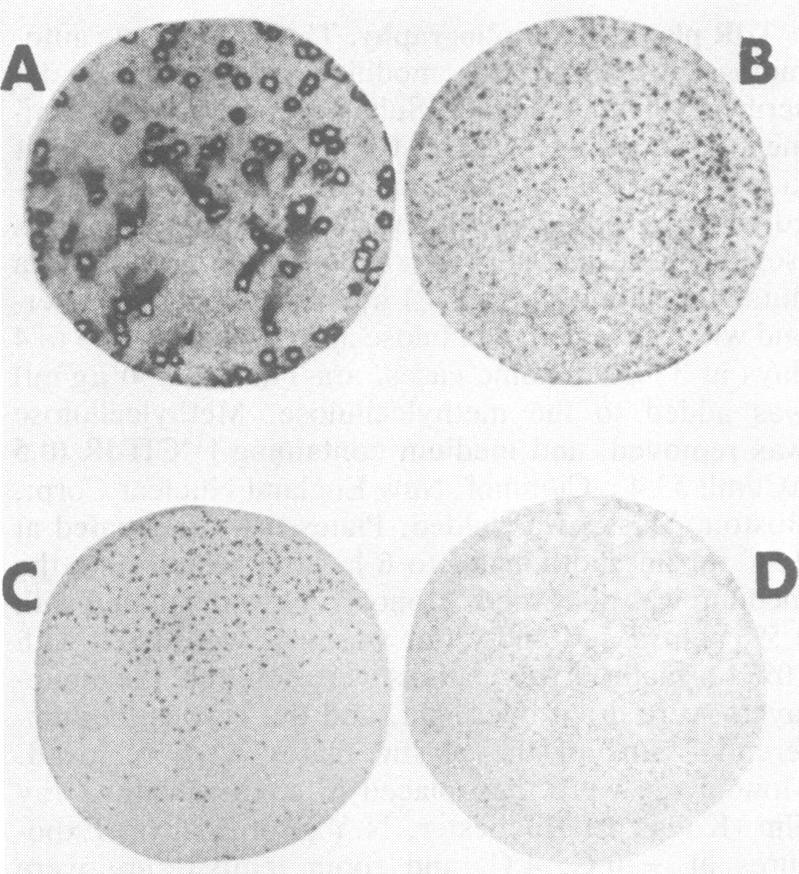
Plaques formed by herpes simplex virus (HSV), pseudorabies virus, and varicella-zoster virus were studied by plaque autoradiography after [14C]thymidine labeling. Standard thymidine kinase-positive (TK+) viruses and TK- mutants of HSV types 1 and 2 and pseudorabies virus were studied, including cell cultured viruses and viruses isolated from animals. Autoradiography was performed with X-ray film with an exposure time of 5 days. After development of films, TK+ plaques showed dark rims due to isotope incorporation, whereas TK- plaques were minimally labeled. Plaque autoradiography of stock TK- viruses showed reversion frequencies to the TK+ phenotype of less than 10(-3). Autoradiography indicated that TK- virus retained the TK- phenotype after replication in vivo. In addition, it was shown that TK- HSV could be isolated from mouse trigeminal ganglion tissue after corneal inoculation of TK- HSV together with TK+ HSV. The plaque autoradiographic procedure was very useful to evaluate proportions of TK+ and TK- virus present in TK+-TK- virus mixtures.
在[14C]胸苷标记后,通过噬斑放射自显影术研究了由单纯疱疹病毒(HSV)、伪狂犬病病毒和水痘-带状疱疹病毒形成的噬斑。研究了1型和2型HSV以及伪狂犬病病毒的标准胸苷激酶阳性(TK+)病毒和TK-突变体,包括细胞培养病毒和从动物体内分离的病毒。使用X射线胶片进行放射自显影,曝光时间为5天。胶片显影后,由于同位素掺入,TK+噬斑显示出深色边缘,而TK-噬斑标记极少。库存TK-病毒的噬斑放射自显影显示,其向TK+表型的回复频率低于10(-3)。放射自显影表明,TK-病毒在体内复制后仍保留TK-表型。此外,研究表明,在将TK- HSV与TK+ HSV一起角膜接种后,可从小鼠三叉神经节组织中分离出TK- HSV。噬斑放射自显影程序对于评估TK+-TK-病毒混合物中TK+和TK-病毒的比例非常有用。