• 文献检索
  • 文档翻译
  • 深度研究
  • 学术资讯
  • Suppr Zotero 插件Zotero 插件
  • 邀请有礼
  • 套餐&价格
  • 历史记录
应用&插件
Suppr Zotero 插件Zotero 插件浏览器插件Mac 客户端Windows 客户端微信小程序
定价
高级版会员购买积分包购买API积分包
服务
文献检索文档翻译深度研究API 文档MCP 服务
关于我们
关于 Suppr公司介绍联系我们用户协议隐私条款
关注我们

Suppr 超能文献

核心技术专利:CN118964589B侵权必究
粤ICP备2023148730 号-1Suppr @ 2026

文献检索

告别复杂PubMed语法,用中文像聊天一样搜索,搜遍4000万医学文献。AI智能推荐,让科研检索更轻松。

立即免费搜索

文件翻译

保留排版,准确专业,支持PDF/Word/PPT等文件格式,支持 12+语言互译。

免费翻译文档

深度研究

AI帮你快速写综述,25分钟生成高质量综述,智能提取关键信息,辅助科研写作。

立即免费体验

体外超声照射细胞的诊断超声延时及透射电子显微镜研究

Diagnostic ultrasound time-lapse and transmission electron microscopic studies of cells insonated in vitro.

作者信息

Liebeskind D, Padawer J, Wolley R, Bases R

出版信息

Br J Cancer Suppl. 1982 Mar;5:176-86.

PMID:6950757
原文链接:https://pmc.ncbi.nlm.nih.gov/articles/PMC2149293/
Abstract

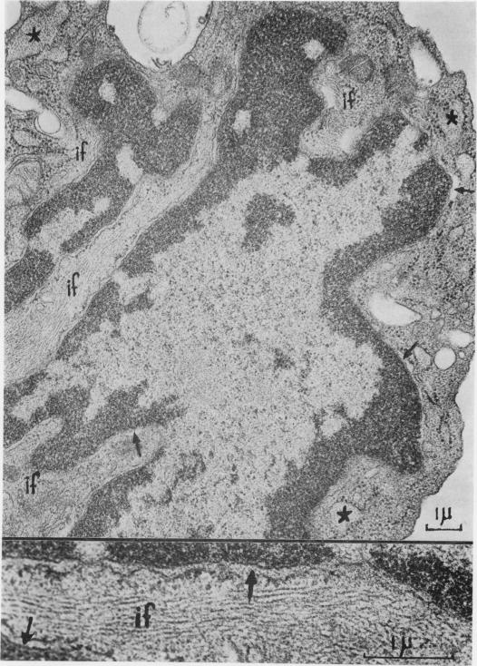
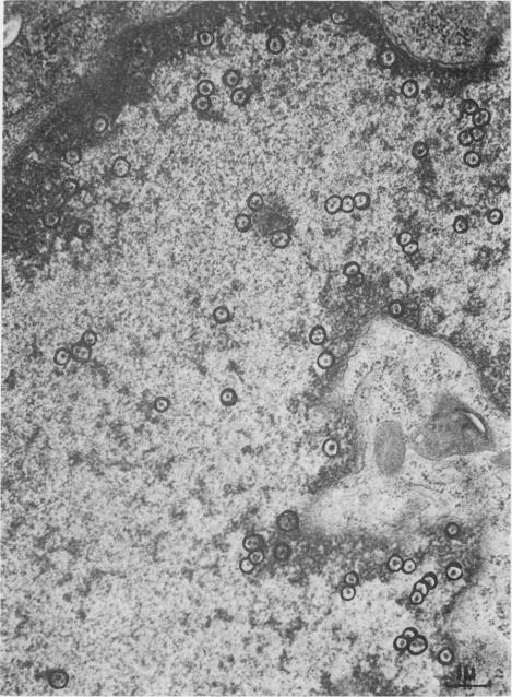
A fibroblast cell line (3T3) and normal rat peritoneal fluid cells were exposed in vitro to pulsed ultrasound from a diagnostic instrument (Smith-Kline "Ekoline 20"). We report here on ultrastructural changes in both cell types and on altered motility patterns in 3T3 fibroblasts. Abnormal motility was detectable 10 generations after exposure. X-irradiation and ultraviolet light elicited similar effects on cell motion. It is suggested that the cellular effects of diagnostic levels of ultrasound be further examined both in vitro and in vivo.

摘要

将成纤维细胞系(3T3)和正常大鼠腹腔液细胞在体外暴露于一台诊断仪器(史克必成公司的“Ekoline 20”)发出的脉冲超声。我们在此报告这两种细胞类型的超微结构变化以及3T3成纤维细胞运动模式的改变。暴露后10代可检测到异常运动。X射线照射和紫外线对细胞运动产生了类似影响。建议在体外和体内进一步研究诊断水平超声的细胞效应。

https://cdn.ncbi.nlm.nih.gov/pmc/blobs/7e9c/2149293/871d3f0acdcf/brjcancersuppl00063-0193-a.jpg
https://cdn.ncbi.nlm.nih.gov/pmc/blobs/7e9c/2149293/d546f24fe4dd/brjcancersuppl00063-0189-a.jpg
https://cdn.ncbi.nlm.nih.gov/pmc/blobs/7e9c/2149293/9564eb2d1b84/brjcancersuppl00063-0186-a.jpg
https://cdn.ncbi.nlm.nih.gov/pmc/blobs/7e9c/2149293/f404cd9e4684/brjcancersuppl00063-0187-a.jpg
https://cdn.ncbi.nlm.nih.gov/pmc/blobs/7e9c/2149293/907eea58f8d3/brjcancersuppl00063-0188-a.jpg
https://cdn.ncbi.nlm.nih.gov/pmc/blobs/7e9c/2149293/ecf445f8af77/brjcancersuppl00063-0190-a.jpg
https://cdn.ncbi.nlm.nih.gov/pmc/blobs/7e9c/2149293/a5eef336c77b/brjcancersuppl00063-0191-a.jpg
https://cdn.ncbi.nlm.nih.gov/pmc/blobs/7e9c/2149293/1d0251809b55/brjcancersuppl00063-0192-a.jpg
https://cdn.ncbi.nlm.nih.gov/pmc/blobs/7e9c/2149293/871d3f0acdcf/brjcancersuppl00063-0193-a.jpg

相似文献

1
Diagnostic ultrasound time-lapse and transmission electron microscopic studies of cells insonated in vitro.体外超声照射细胞的诊断超声延时及透射电子显微镜研究
Br J Cancer Suppl. 1982 Mar;5:176-86.
2
NTP technical report on the toxicity studies of Dibutyl Phthalate (CAS No. 84-74-2) Administered in Feed to F344/N Rats and B6C3F1 Mice.美国国家毒理学计划关于邻苯二甲酸二丁酯(化学物质登记号84 - 74 - 2)经饲料给予F344/N大鼠和B6C3F1小鼠的毒性研究技术报告。
Toxic Rep Ser. 1995 Apr;30:1-G5.
3
Centrioles, microtubules and microfilaments in activated mononuclear and multinucleate macrophages from rat peritoneum: electron-microscopic and immunofluorescence microscopic studies.大鼠腹膜活化单核及多核巨噬细胞中的中心粒、微管和微丝:电子显微镜及免疫荧光显微镜研究
J Pathol. 1981 Apr;133(4):301-23. doi: 10.1002/path.1711330404.
4
The response of megakaryocytes with processes to thrombin.有突起的巨核细胞对凝血酶的反应。
Thromb Haemost. 1987 Aug 4;58(2):732-6.
5
[Structural change in the cell center during fibroblast polarization and movement].[成纤维细胞极化和迁移过程中细胞中心的结构变化]
Tsitologiia. 1983 Aug;25(8):883-8.
6
Plate-induced tumors of BALB/3T3 cells exhibiting foci of differentiation into pericytes, chondrocytes, and fibroblasts.平板诱导的BALB/3T3细胞肿瘤,表现出向周细胞、软骨细胞和成纤维细胞分化的灶状区域。
J Supramol Struct. 1980;14(2):233-40. doi: 10.1002/jss.400140212.
7
A study of the influence of the ambient microflora on the structure of lung alveolar macrophages and an ultrastructural comparison of lung and peritoneal macrophages in germ-free and conventionally reared mice.无菌饲养和常规饲养小鼠中环境微生物群对肺泡巨噬细胞结构的影响以及肺巨噬细胞与腹腔巨噬细胞的超微结构比较研究。
J Morphol. 1981 Sep;169(3):283-91. doi: 10.1002/jmor.1051690304.
8
[Cell center of macrophages, granulocytes and lymphocytes during in vitro cell spreading, polarization and movement].[体外细胞铺展、极化和运动过程中巨噬细胞、粒细胞和淋巴细胞的细胞中心]
Tsitologiia. 1984 Sep;26(9):1002-7.
9
Regulation of the functional activity of mast cells and fibroblasts by mononuclear cells in murine and human chronic graft-vs.-host disease.单核细胞对小鼠和人类慢性移植物抗宿主病中肥大细胞和成纤维细胞功能活性的调节
Exp Hematol. 1997 Mar;25(3):238-45.
10
The fine structure of macrophage granules in experimental granulomas in rodents.啮齿动物实验性肉芽肿中巨噬细胞颗粒的精细结构。
J Anat. 1979 May;128(Pt 3):479-87.

引用本文的文献

1
Section 8--clinical relevance. American Institute of Ultrasound in Medicine.第8节——临床相关性。美国医学超声学会。
J Ultrasound Med. 2000 Feb;19(2):149-68. doi: 10.7863/jum.2000.19.2.149.
2
Section 7--discussion of the mechanical index and other exposure parameters. American Institute of Ultrasound in Medicine.第7节——机械指数及其他暴露参数的讨论。美国医学超声学会。
J Ultrasound Med. 2000 Feb;19(2):143-8, 154-68. doi: 10.7863/jum.2000.19.2.143.
3
Section 6--mechanical bioeffects in the presence of gas-carrier ultrasound contrast agents. American Institute of Ultrasound in Medicine.

本文引用的文献

1
Morphological changes in the surface characteristics of cultured cells after exposure to diagnostic ultrasound.暴露于诊断超声后培养细胞表面特征的形态学变化。
Radiology. 1981 Feb;138(2):419-23. doi: 10.1148/radiology.138.2.7455124.
2
Functional significance of perichromatin granule accumulation induced by cadmium chloride in isolated rat liver cells.氯化镉诱导分离的大鼠肝细胞中染色质外周颗粒积累的功能意义。
Exp Cell Res. 1980 Jul;128(1):47-58. doi: 10.1016/0014-4827(80)90385-7.
3
Has diagnostic ultrasound mutagenic effects?诊断性超声有诱变作用吗?
第6节——气体载体超声造影剂存在下的机械生物效应。美国超声医学学会。
J Ultrasound Med. 2000 Feb;19(2):120-42, 154-68. doi: 10.7863/jum.2000.19.2.120.
4
Section 4--bioeffects in tissues with gas bodies. American Institute of Ultrasound in Medicine.第4节——含气体组织中的生物效应。美国医学超声学会。
J Ultrasound Med. 2000 Feb;19(2):97-108, 154-68. doi: 10.7863/jum.2000.19.2.97.
5
Routine ultrasonography in utero and subsequent handedness and neurological development.子宫内常规超声检查及随后的利手和神经发育
BMJ. 1993 Jul 17;307(6897):159-64. doi: 10.1136/bmj.307.6897.159.
Hum Genet. 1980;56(1):95-8. doi: 10.1007/BF00281576.
4
Relationship between x-ray exposure and malignant transformation in C3H 10T1/2 cells.C3H 10T1/2细胞中X射线照射与恶性转化的关系。
Proc Natl Acad Sci U S A. 1980 Dec;77(12):7262-6. doi: 10.1073/pnas.77.12.7262.
5
Nuclear ultrastructure of epithelial cell lines derived from human carcinomas and nonmalignant tissues.
Cancer Res. 1979 Feb;39(2 Pt 1):332-44.
6
Sister chromatid exchanges in human lymphocytes after exposure to diagnostic ultrasound.诊断性超声暴露后人淋巴细胞中的姐妹染色单体交换
Science. 1979 Sep 21;205(4412):1273-5. doi: 10.1126/science.472742.
7
Diagnostic ultrasound: effects on the DNA and growth patterns of animal cells.诊断超声:对动物细胞DNA和生长模式的影响
Radiology. 1979 Apr;131(1):177-84. doi: 10.1148/131.1.177.