• 文献检索
  • 文档翻译
  • 深度研究
  • 学术资讯
  • Suppr Zotero 插件Zotero 插件
  • 邀请有礼
  • 套餐&价格
  • 历史记录
应用&插件
Suppr Zotero 插件Zotero 插件浏览器插件Mac 客户端Windows 客户端微信小程序
定价
高级版会员购买积分包购买API积分包
服务
文献检索文档翻译深度研究API 文档MCP 服务
关于我们
关于 Suppr公司介绍联系我们用户协议隐私条款
关注我们

Suppr 超能文献

核心技术专利:CN118964589B侵权必究
粤ICP备2023148730 号-1Suppr @ 2026

文献检索

告别复杂PubMed语法,用中文像聊天一样搜索,搜遍4000万医学文献。AI智能推荐,让科研检索更轻松。

立即免费搜索

文件翻译

保留排版,准确专业,支持PDF/Word/PPT等文件格式,支持 12+语言互译。

免费翻译文档

深度研究

AI帮你快速写综述,25分钟生成高质量综述,智能提取关键信息,辅助科研写作。

立即免费体验

The formation, septation and fate of the truncus arteriosus in man.

作者信息

Orts-Llorca F, Puerta Fonolla J, Sobrado J

出版信息

J Anat. 1982 Jan;134(Pt 1):41-56.

PMID:7076544
原文链接:https://pmc.ncbi.nlm.nih.gov/articles/PMC1167935/
Abstract

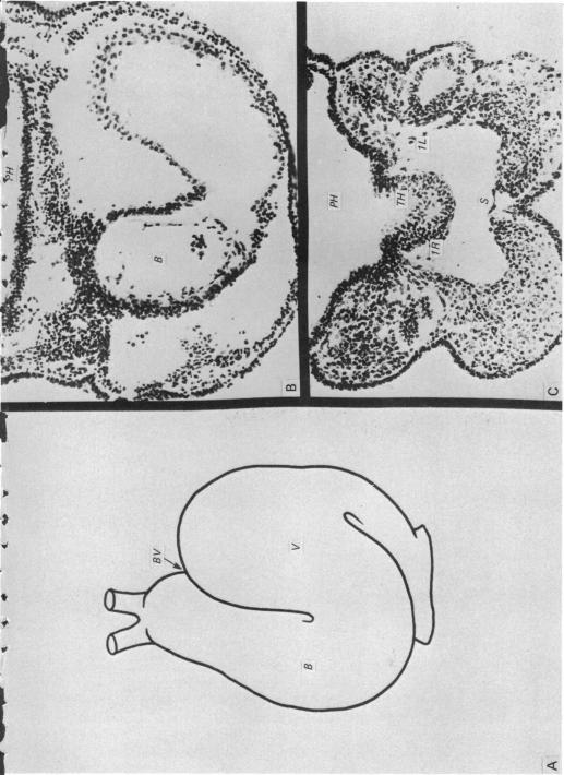
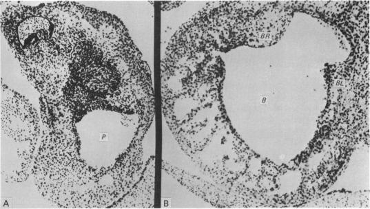
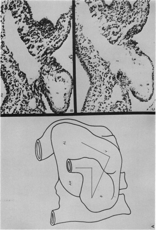
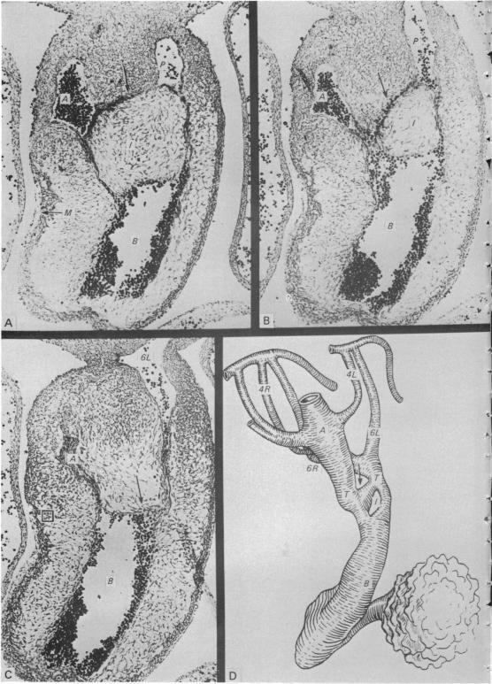
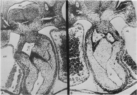
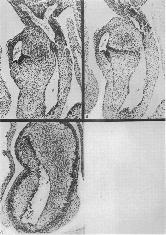
The results of our studies enable us to draw the following conclusions. The truncus appears in the human embryo, between Stages XII and XIII, as a portion of the aortic sac which invaginates into the interior of the pericardial cavity. Therefore it is an arterial portion which is added to the heart. It lengthens progressively. The sigmoid valves form in the angle between the bulbus cordis and the truncus. Septation of the truncus begins when the sixth arterial arches appear in embryos of 6 to 8 mm. The process is very rapid; commencing in embryos of 6 to 7 mm, it is complete in embryos of 10 to 11 mm, that is to say, during only five days. The septation mechanism is extrinsic. The peribranchial mesenchyma which accompanies the aortic sac in its invagination advances principally on the right inferior part and insinuates itself between the fourth and sixth arterial arches, separating the truncus pulmonalis from a portion of the ascending aorta. An aorticopulmonary communication exists for a certain period prior to fusion of the two blocks of mesenchyma; there is a mesenchymal island. On the contrary, in the bulbus cordis septation is effected by the bulbar ridges. Septation of the truncus, which does not exist in the primitive cardiac tube, occurs prior to that of the bulbus cordis. While septation of the truncus has been already completed in embryos of 10 mm, septation of the bulbus cordis is completed only in embryos of 14 to 16 mm. Therefore the bulbus and the truncus are two portions, different in respect of both structure and septation. There is no continuity between the bulbar ridges and the septation of the truncus. They are separated by the sigmoid valves. This makes it possible to observe independent malformations in the bulbus and in the truncus. In the truncus the mesenchyma passes between the two vessels. They do not have a common septum, and it is for this reason that the surgeon can separate them in the mature heart.

摘要
https://cdn.ncbi.nlm.nih.gov/pmc/blobs/d213/1167935/24d8bc98bdfd/janat00217-0058-a.jpg
https://cdn.ncbi.nlm.nih.gov/pmc/blobs/d213/1167935/e1e91fd7492d/janat00217-0050-a.jpg
https://cdn.ncbi.nlm.nih.gov/pmc/blobs/d213/1167935/6124df2d90a6/janat00217-0051-a.jpg
https://cdn.ncbi.nlm.nih.gov/pmc/blobs/d213/1167935/d6e8ce9a5716/janat00217-0053-a.jpg
https://cdn.ncbi.nlm.nih.gov/pmc/blobs/d213/1167935/824d6545d159/janat00217-0055-a.jpg
https://cdn.ncbi.nlm.nih.gov/pmc/blobs/d213/1167935/92a93b95e04e/janat00217-0056-a.jpg
https://cdn.ncbi.nlm.nih.gov/pmc/blobs/d213/1167935/fb167c6d0dbb/janat00217-0057-a.jpg
https://cdn.ncbi.nlm.nih.gov/pmc/blobs/d213/1167935/24d8bc98bdfd/janat00217-0058-a.jpg

相似文献

1
The formation, septation and fate of the truncus arteriosus in man.
J Anat. 1982 Jan;134(Pt 1):41-56.
2
The septation of the arterial pole of the heart in the chick embryo. III. Development of the truncus arteriosus of the heart of chick embryos from 5 1/2 to 7 days of incubation.鸡胚心脏动脉极的分隔。III. 孵化5.5至7天鸡胚心脏动脉干的发育。
Acta Morphol Neerl Scand. 1979 Feb;17(1):1-20.
3
Histological study of the proximal and distal segments of the embryonic outflow tract and great arteries.胚胎流出道和大动脉近端及远端节段的组织学研究。
Anat Rec A Discov Mol Cell Evol Biol. 2005 Mar;283(1):202-11. doi: 10.1002/ar.a.20138.
4
Morphogenesis of the truncus arteriosus of the chick embryo heart: tissue reorganization during septation.鸡胚心脏动脉干的形态发生:分隔过程中的组织重组。
Am J Anat. 1979 Oct;156(2):251-64. doi: 10.1002/aja.1001560206.
5
Morphogenesis of the truncus arteriosus of the chick embryo heart: movements of autoradiographic tattoos during septation.
Anat Rec. 1987 Aug;218(4):434-40, 394-5. doi: 10.1002/ar.1092180411.
6
Morphogenesis of the truncus arteriosus of the chick embryo heart: the formation and migration of mesenchymal tissue.鸡胚心脏动脉干的形态发生:间充质组织的形成与迁移。
Am J Anat. 1979 Apr;154(4):545-56. doi: 10.1002/aja.1001540408.
7
Experimental study of the development of the truncus and the conus in the chick embryo.
J Anat. 1977 Jul;123(Pt 3):661-86.
8
Experimental study of the development of the truncus arteriosus of the chick embryo heart. I. Time of appearance.鸡胚心脏动脉干发育的实验研究。I. 出现时间。
Anat Rec. 1993 Nov;237(3):378-84. doi: 10.1002/ar.1092370311.
9
Development of the musculoelastic septation complex in the avian truncus arteriosus.鸟类动脉干中肌弹性分隔复合体的发育
Am J Anat. 1990 Dec;189(4):339-56. doi: 10.1002/aja.1001890406.
10
The septation of the arterial pole of the heart in the chick embryo. II. Development of the truncus arteriosus of the heart of chick embryos from 4 to 5 days of incubation.
Acta Morphol Neerl Scand. 1978 Feb;16(1):29-53 contd.

引用本文的文献

1
Human Cardiac Development.人类心脏发育
Adv Exp Med Biol. 2024;1441:3-55. doi: 10.1007/978-3-031-44087-8_1.
2
Spatially Distinct Genetic Determinants of Aortic Dimensions Influence Risks of Aneurysm and Stenosis.主动脉尺寸的空间差异遗传决定因素影响动脉瘤和狭窄的风险。
J Am Coll Cardiol. 2022 Aug 2;80(5):486-497. doi: 10.1016/j.jacc.2022.05.024.
3
A pictorial account of the human embryonic heart between 3.5 and 8 weeks of development.人类胚胎心脏在 3.5 至 8 周发育期间的图示描述。

本文引用的文献

1
Comparative development of the bulbus and ventricles of the vertebrate heart with special reference to Spitzer's theory of heart malformations.
Anat Rec. 1962 Apr;142:519-29. doi: 10.1002/ar.1091420409.
2
[Development of the septum sinus venosi cordis. Development of the heart in man, determined from a forgotten structure].
Z Anat Entwicklungsgesch. 1960;122:173-96.
3
[The development of the interventricular syptum in the human heart, with special reference to bulbus-torsion].[人类心脏室间隔的发育,特别提及球室扭转]
Z Anat Entwicklungsgesch. 1969;128(1):1-17.
4
Commun Biol. 2022 Mar 11;5(1):226. doi: 10.1038/s42003-022-03153-x.
4
Development of the Human Arterial Valves: Understanding Bicuspid Aortic Valve.人类动脉瓣膜的发育:了解二叶式主动脉瓣
Front Cardiovasc Med. 2022 Jan 27;8:802930. doi: 10.3389/fcvm.2021.802930. eCollection 2021.
5
The proximal segment of the embryonic outflow (conus) does not participate in aortic vestibule development.胚胎流出道(圆锥)的近端部分不参与主动脉前庭的发育。
PLoS One. 2018 Dec 31;13(12):e0209930. doi: 10.1371/journal.pone.0209930. eCollection 2018.
6
Developmental Progression of the Coronary Vasculature in Human Embryos and Fetuses.人类胚胎和胎儿冠状动脉血管的发育进程
Anat Rec (Hoboken). 2016 Jan;299(1):25-41. doi: 10.1002/ar.23283. Epub 2015 Nov 28.
7
A detailed comparison of mouse and human cardiac development.小鼠与人类心脏发育的详细比较。
Pediatr Res. 2014 Dec;76(6):500-7. doi: 10.1038/pr.2014.128. Epub 2014 Aug 28.
8
Normal and abnormal development of the intrapericardial arterial trunks in humans and mice.人类和小鼠的心包内动脉干的正常和异常发育。
Cardiovasc Res. 2012 Jul 1;95(1):108-15. doi: 10.1093/cvr/cvs147. Epub 2012 Apr 12.
9
Three-dimensional and molecular analysis of the arterial pole of the developing human heart.人类心脏动脉干的三维和分子分析。
J Anat. 2012 Apr;220(4):336-49. doi: 10.1111/j.1469-7580.2012.01474.x. Epub 2012 Feb 1.
10
Phylogeny informs ontogeny: a proposed common theme in the arterial pole of the vertebrate heart.系统发生启示个体发生:脊椎动物心脏动脉极中的一个拟议的共同主题。
Evol Dev. 2010 Nov-Dec;12(6):552-67. doi: 10.1111/j.1525-142X.2010.00441.x.
The conotruncus. I. Its normal inversion and conus absorption.
Circulation. 1972 Aug;46(2):375-84. doi: 10.1161/01.cir.46.2.375.
5
Anatomical study of truncus arteriousus communis with embryological and surgical considerations.共同动脉干的解剖学研究及胚胎学与手术学考量
Br Heart J. 1976 Nov;38(11):1109-23. doi: 10.1136/hrt.38.11.1109.
6
Experimental study of the development of the truncus and the conus in the chick embryo.
J Anat. 1977 Jul;123(Pt 3):661-86.
7
Development of the outflow tract of the embryonic heart.
Birth Defects Orig Artic Ser. 1978;14(7):29-68.
8
Cardiac septation and development of the aorta, pulmonary trunk, and pulmonary veins: previous work in the light of recent observations.
Birth Defects Orig Artic Ser. 1978;14(7):109-38.
9
Morphogenesis of congenital heart anomaly--bulboventricular malformations.
Jpn Circ J. 1978 Oct;42(10):1105-20. doi: 10.1253/jcj.42.1105.
10
Scanning and light microscope studies of the development of the chick embryo semilunar heart valves.
Anat Embryol (Berl). 1979;157(1):69-80. doi: 10.1007/BF00315641.