Coddington R, Mera S L, Goddard P R, Bradfield J W
J Clin Pathol. 1982 May;35(5):536-40. doi: 10.1136/jcp.35.5.536.
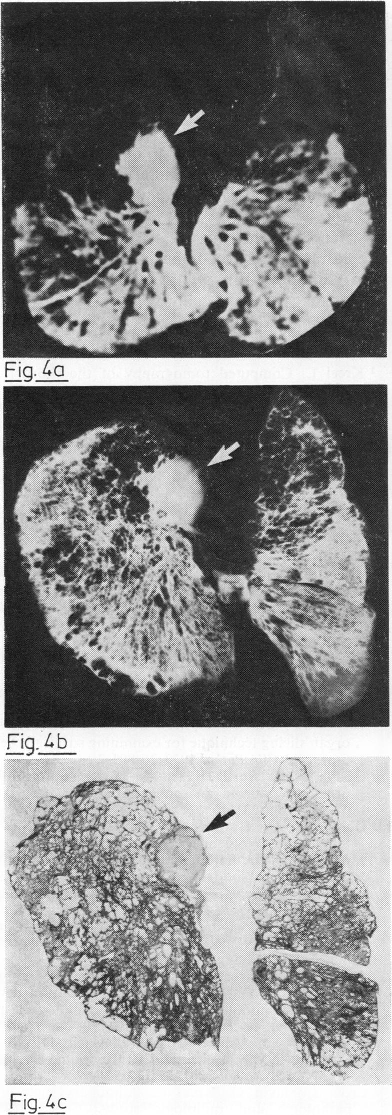
A method is described which allows the features seen in images generated during computed tomography (CT) of lungs previously removed at necropsy to be compared with those seen in corresponding thin sections made from the same lungs. After removal from the body, lungs were inflation-fixed using warm formalin vapour before being x-rayed and then scanned in the inflated state. Slices corresponding to the CT scan images were made and x-rayed. Paper mounted sections were then prepared from each slice. Using these methods pathological correlative studies can be used both to validate the interpretation of CT scans of lungs and to assess the sensitivity of this imaging technique.
本文描述了一种方法,该方法可将先前尸检时切除的肺部在计算机断层扫描(CT)中生成的图像特征与取自同一肺部的相应薄片中的特征进行比较。从身体取出后,肺部先用温热的福尔马林蒸汽进行充气固定,然后进行X光检查,接着在充气状态下进行扫描。制作与CT扫描图像对应的切片并进行X光检查。然后从每个切片制备裱贴在纸上的切片。使用这些方法,病理相关性研究既可以用于验证肺部CT扫描的解读,也可以用于评估这种成像技术的敏感性。