Sone S, Bucana C, Hoyer L C, Fidler I J
Am J Pathol. 1981 May;103(2):234-46.
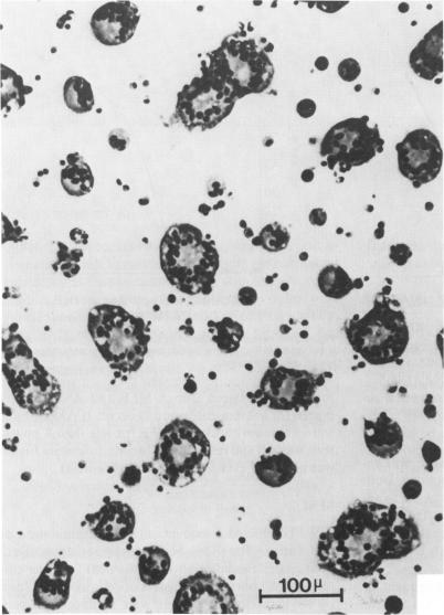
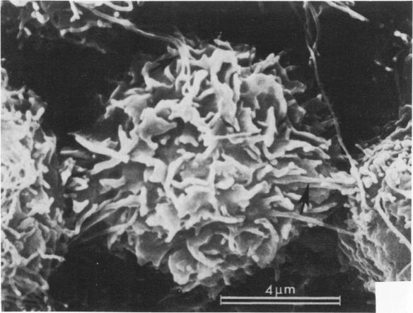
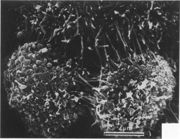
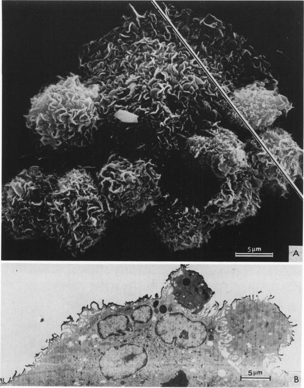
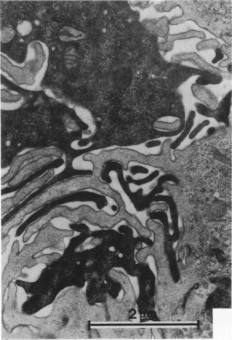
Treatment of F344 rat alveolar macrophages (AMs) in vitro with cell-free supernatant fluids obtained from concanavalin-A (Con A)-stimulated syngeneic lymphocytes induced extensive fusion. The lymphokine responsible for the fusion of AMs (but not other cells) is here referred to as AM fusion factor (Con-A-MFF). Fusion is dependent on the dose of Con-A-MFF and the population density of AM cultures and occurred 10 hours after Con-A-MFF was added to cultures of normal AMs. Con-A-MFF must interact with AMs for more than 8 hours before full expression of fusion is reached at 24 hours. Using a technique allowing for sequential scanning to transmission electron microscopy analysis of cells, the authors determined the relationship of the morphologic characteristics of the surface and the internal structure of cells fusing to form multinucleate giant cells (MGCs). The process of AM fusion begins with the aggregation of AMs, followed by interdigitation of cell processes. Serial sections of MGCs showed lysosomes associated with remnants of plasma membrane in the cytoplasm. The MGCs contained numerous organelles associated with increased secretory activity of cells.
用从刀豆蛋白A(Con A)刺激的同基因淋巴细胞获得的无细胞上清液体外处理F344大鼠肺泡巨噬细胞(AMs),可诱导广泛融合。负责AMs(而非其他细胞)融合的淋巴因子在此被称为AM融合因子(Con-A-MFF)。融合取决于Con-A-MFF的剂量和AM培养物的细胞群体密度,在将Con-A-MFF添加到正常AMs培养物后10小时发生。Con-A-MFF必须与AMs相互作用超过8小时,才能在24小时达到融合的充分表达。作者使用一种允许对细胞进行连续扫描以进行透射电子显微镜分析的技术,确定了融合形成多核巨细胞(MGCs)的细胞表面形态特征与内部结构之间的关系。AMs融合过程始于AMs的聚集,随后是细胞突起的相互交错。MGCs的连续切片显示溶酶体与细胞质中的质膜残余物相关。MGCs含有许多与细胞分泌活性增加相关的细胞器。