Pu L P, Van Leeuwen F W, Tracer H L, Sonnemans M A, Loh Y P
Section on Cellular Neurobiology, National Institute of Child Health and Human Development, National Institutes of Health, Bethesda, MD 20892, USA.
Proc Natl Acad Sci U S A. 1995 Nov 7;92(23):10653-7. doi: 10.1073/pnas.92.23.10653.
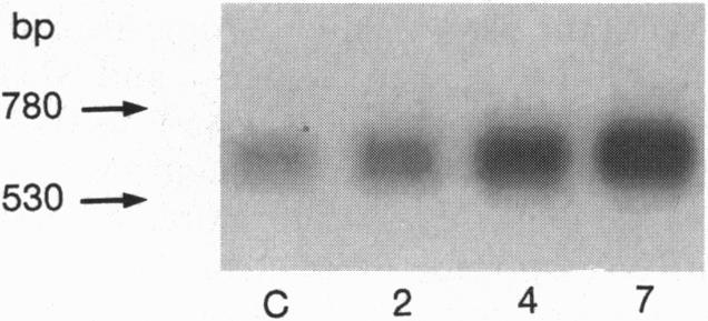
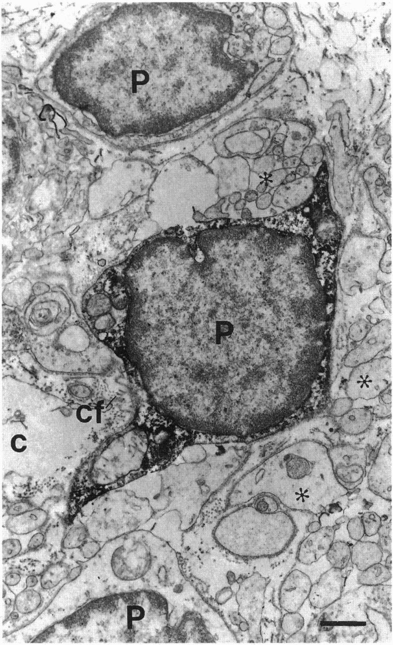
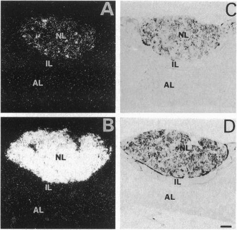
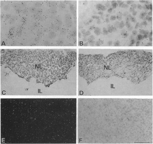
The presence of [arginine] vasopressin (AVP) mRNA and AVP immunoreactivity in pituicytes of the neural lobe (NL) of intact and pituitary stalk-transected rats, with and without osmotic stimulation, was examined. AVP mRNA was analyzed by Northern blotting, as well as by in situ hybridization in combination with immunocytochemistry using anti-glial fibrillary acidic protein (GFAP) as a marker for pituicytes. In intact rats, a poly(A) tail-truncated 0.62-kb AVP mRNA was detected in the NL and was found to increase 10-fold with 7 days of continuous salt loading. Morphological analysis of the NL of 7-day salt-loaded rats revealed the presence of AVP mRNA in a significant number of GFAP-positive pituicytes in the NL and in areas most probably containing nerve fibers. Eight days after pituitary stalk transection the NL AVP mRNA diminished in animals given water to drink, whereas in those given 2% saline for 18 h followed by 6 h of water, a treatment repeated on 6 successive days beginning 2 days after surgery, the 0.62-kb AVP mRNA was present. The AVP mRNA in the pituitary stalk-transected, salt-loaded rats showed an exclusive cellular distribution in the NL, indicative of localization in pituicytes. Immunoelectron microscopy showed the presence of AVP immunoreactivity in a subpopulation of pituicytes 7 and 10 days after pituitary stalk transection in salt-loaded animals, when almost all AVP fibers had disappeared from the NL. These data show that a subset of pituicytes in the NL is activated to synthesize AVP mRNA and AVP in response to osmotic stimulation.
对完整的和垂体柄横断的大鼠神经垂体(NL)的垂体细胞中[精氨酸]血管加压素(AVP)mRNA和AVP免疫反应性进行了检测,检测时伴有或不伴有渗透刺激。通过Northern印迹法以及原位杂交结合免疫细胞化学分析AVP mRNA,免疫细胞化学使用抗胶质纤维酸性蛋白(GFAP)作为垂体细胞的标志物。在完整大鼠中,在神经垂体中检测到一种多聚腺苷酸尾截短的0.62-kb AVP mRNA,并且发现连续7天给予高盐负荷后其增加了10倍。对7天高盐负荷大鼠的神经垂体进行形态学分析,发现在神经垂体中大量GFAP阳性垂体细胞以及很可能含有神经纤维的区域存在AVP mRNA。垂体柄横断8天后,给予饮水的动物神经垂体AVP mRNA减少,而给予2%盐水18小时然后给予6小时水的动物(从手术后2天开始连续6天重复这种处理),存在0.62-kb AVP mRNA。垂体柄横断且高盐负荷大鼠中的AVP mRNA在神经垂体中呈现独特的细胞分布,表明其定位于垂体细胞。免疫电子显微镜显示,在高盐负荷动物垂体柄横断7天和10天后,垂体细胞亚群中存在AVP免疫反应性,此时几乎所有AVP纤维已从神经垂体消失。这些数据表明,神经垂体中的一部分垂体细胞在受到渗透刺激时被激活以合成AVP mRNA和AVP。