Berthon P, Dimitrov T, Stower M, Cussenot O, Maitland N J
Department of Biology, University of York, Heslington, UK.
Br J Cancer. 1995 Oct;72(4):946-51. doi: 10.1038/bjc.1995.439.
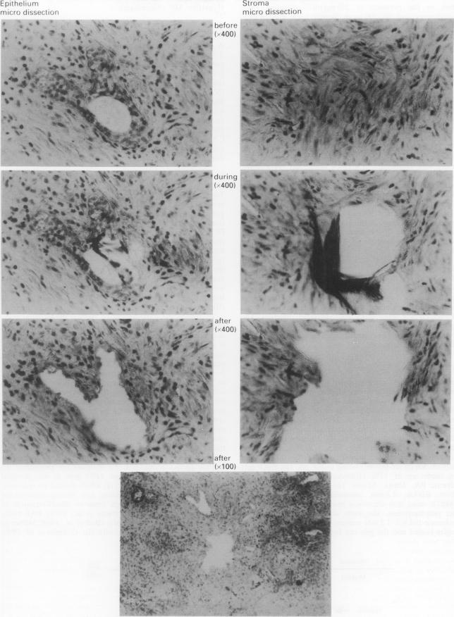
To investigate the underlying mechanisms of carcinogenesis, we have developed a technique to determine the frequency of genetic changes in prostatic carcinoma tissue. We have demonstrated that at a ratio of between 1:4 and 1:9 mutant-normal alleles, the signal from a mutant TP53 allele is not apparent after polymerase chain reaction (PCR) amplification and further direct sequencing or single-strand conformation polymorphism (SSCP) analysis. To bypass this problem, which is inherent in the heterogeneity of the prostate tissue and of the tumour, we selected areas of graded prostate tumours (Gleason score) from cryosectioned preparations and microdissected these cells (20-100 cells). After anionic resin removal of proteins, PCR amplification of TP53 gene exons 5/6 and SSCP analysis, an abnormal SSCP band shift was observed in suspected tumour cells, compared with microdissected stromal cells used as an internal control, while (1) a crude preparation of tissue DNA carrying the tumour did not show any abnormality and (2) immunostaining by a set of monoclonal antibodies against TP53 protein remained negative. Nucleotide sequence analysis of the different bands confirmed the presence of a mutation in the TP53 gene exon 6 position 13,336 in an abnormal band for one specimen, while no mutation was detected in the normal SSCP band. By targeting recognised tumour cells we can find DNA mutations which are undetectable using the standard technique of whole-tissue DNA extraction, particularly in a heterogeneous tumour such as carcinoma of the prostate.
为了研究致癌作用的潜在机制,我们开发了一种技术来确定前列腺癌组织中基因变化的频率。我们已经证明,当突变型与正常等位基因的比例在1:4至1:9之间时,聚合酶链反应(PCR)扩增以及进一步的直接测序或单链构象多态性(SSCP)分析后,来自突变型TP53等位基因的信号并不明显。为了绕过这个前列腺组织和肿瘤异质性所固有的问题,我们从冷冻切片标本中选择了不同分级的前列腺肿瘤区域(Gleason评分),并对这些细胞进行显微切割(20 - 100个细胞)。在用阴离子树脂去除蛋白质后,对TP53基因第5/6外显子进行PCR扩增和SSCP分析,与用作内部对照的显微切割基质细胞相比,在疑似肿瘤细胞中观察到异常的SSCP条带迁移,而(1)携带肿瘤的组织DNA粗提物未显示任何异常,并且(2)用一组抗TP53蛋白的单克隆抗体进行免疫染色仍为阴性。对不同条带的核苷酸序列分析证实,一个标本的异常条带中TP53基因第6外显子13336位存在突变,而在正常SSCP条带中未检测到突变。通过靶向识别肿瘤细胞,我们能够发现使用全组织DNA提取的标准技术无法检测到的DNA突变,特别是在前列腺癌这种异质性肿瘤中。