Lawrenson J G, Ghabriel M N, Reid A R, Gajree T N, Allt G
Reta Lila Weston Institute of Neurological Studies, University College London Medical School, UK.
J Anat. 1995 Feb;186 ( Pt 1)(Pt 1):217-21.
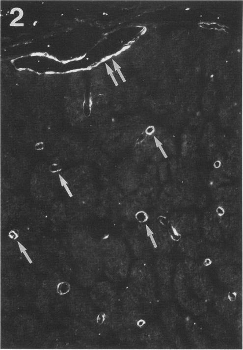
A monoclonal antibody to an antigen (EBA) expressed by neural endothelial cells (EC) was used to investigate any difference in the distribution of EBA between the CNS and PNS. Pre-embedding ultrastructural cytochemistry of rat sciatic and optic nerves was undertaken using anti-EBA, detected with a silver-enhanced gold-conjugated secondary antibody. LM immunocytochemical localisation of EBA was also performed using an HRP-conjugated secondary antibody. EC of pial and parenchymal optic nerve vessels were strongly immunopositive for EBA. Vessels of the dura were negative. At the EM level EBA was observed on the EC luminal surface. In contrast, EC of sciatic nerve were either negative or only weakly immunopositive. The molecular characteristics and function of EBA are largely unknown. Therefore the functional significance of the present findings remains to be determined.
一种针对神经内皮细胞(EC)表达的抗原(EBA)的单克隆抗体被用于研究EBA在中枢神经系统(CNS)和外周神经系统(PNS)之间分布的差异。使用抗EBA对大鼠坐骨神经和视神经进行包埋前超微结构细胞化学分析,并用银增强金偶联二抗进行检测。还使用辣根过氧化物酶(HRP)偶联二抗进行了EBA的光镜免疫细胞化学定位。软脑膜和实质视神经血管的EC对EBA呈强免疫阳性。硬脑膜血管呈阴性。在电镜水平上,EBA出现在EC的管腔表面。相比之下,坐骨神经的EC要么呈阴性,要么仅呈弱阳性免疫反应。EBA的分子特征和功能在很大程度上尚不清楚。因此,目前这些发现的功能意义仍有待确定。