Macdonald A A, Johnstone M
Department of Preclinical Veterinary Sciences, Royal (Dick) School of Veterinary Studies, University of Edinburgh, UK.
J Anat. 1995 Apr;186 ( Pt 2)(Pt 2):235-43.
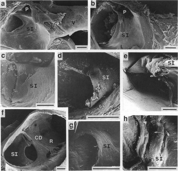
The structure of the foramen ovale from 16 species representing 4 carnivore families, the Felidae, Canidae, Ursidae and Hyaenidae, was studied using the scanning electron microscope. The Felidae were represented by 9 domestic cat fetuses (Felis catus), 2 snow leopard neonates (Uncia uncia), an ocelot neonate (Leopardus pardalis), 2 lion neonates (Panthera leo), a panther neonate (Panthera pardus) and 3 tigers (Neofelis tigris), comprising 2 fetuses and a neonate. The Canidae were represented by a golden jackal neonate (Canis aureus), a newborn wolf (Canis lupus), 8 domestic dog fetuses (Canis familiaris), 3 red fox neonates (Vulpes vulpes) and a dhole neonate (Cuon alpinus). The Ursidae were represented by a brown bear neonate (Ursus arctos), a day-old grizzly bear cub (Ursus arctos horribilis), a polar bear neonate (Ursus maritimus), and 2 additional bear fetuses (species unknown). The Hyaenidae were represented by a striped hyaena neonate (Hyaena hyaena). In each species, the foramen ovale, when viewed from the terminal part of the caudal vena cava, had the appearance of a short tunnel. A thin fold of tissue, the developed remains of the embryonic septum primum, extended from the distal end of the caudal vena cava for a variable distance into the lumen of the left atrium and contributed towards the 'tunnel' appearance in all specimens. It constituted a large proportion of the tube, and its distal end was straight-edged. There was fibrous material underlying the endothelium of the flap, the apparent morphology of which suggested that it comprised cardiac muscle.(ABSTRACT TRUNCATED AT 250 WORDS)
利用扫描电子显微镜研究了代表4个食肉动物科(猫科、犬科、熊科和鬣狗科)的16个物种的卵圆孔结构。猫科动物包括9只家猫胎儿(家猫)、2只雪豹新生儿(雪豹)、1只豹猫新生儿(豹猫)、2只狮子新生儿(狮)、1只黑豹新生儿(豹)和3只老虎(马来貘),其中包括2只胎儿和1只新生儿。犬科动物包括1只金豺新生儿(金豺)、1只新生狼(狼)、8只家犬胎儿(家犬)、3只赤狐新生儿(赤狐)和1只豺新生儿(豺)。熊科动物包括1只棕熊新生儿(棕熊)、1只1日龄灰熊幼崽(灰熊)、1只北极熊新生儿(北极熊)和另外2只熊胎儿(物种未知)。鬣狗科动物包括1只条纹鬣狗新生儿(条纹鬣狗)。在每个物种中,从尾腔静脉末端观察时,卵圆孔呈现出短隧道的外观。一层薄的组织褶皱,即胚胎原发隔的发育残留物,从尾腔静脉远端延伸到左心房腔中一段可变距离,并在所有标本中形成了“隧道”外观。它构成了管道的很大一部分,其远端边缘是直的。瓣叶内皮下方有纤维材料,其明显的形态表明它由心肌组成。(摘要截断于250字)