Van Niekerk C C, Ramaekers F C, Hanselaar A G, Aldeweireldt J, Poels L G
Department of Cell Biology and Histology, Faculty of Medical Sciences, University of Nijmegen, The Netherlands.
Am J Pathol. 1993 Jan;142(1):157-77.
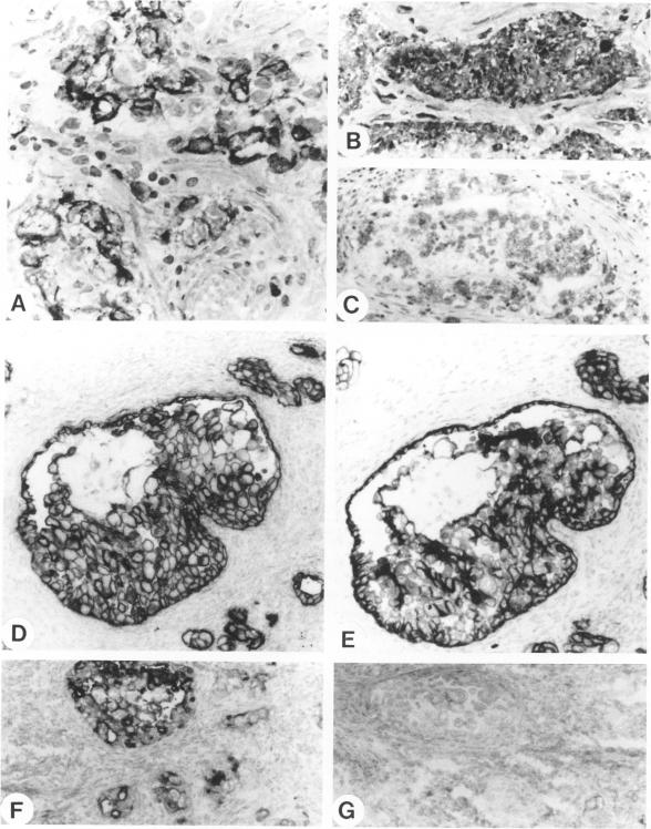
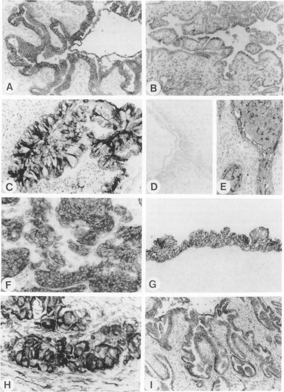
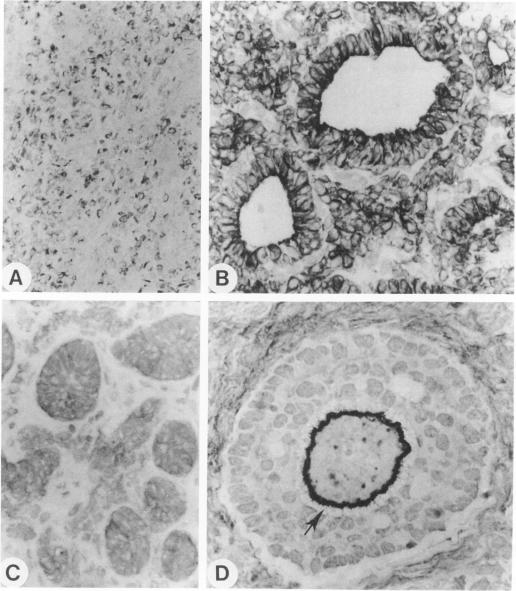
The marker profile of 18 samples of normal human ovarian tissues and 138 samples of their derived tumors was established using 51 monoclonal antibodies directed against intermediate filaments, ovarian carcinoma-specific antigens, general tumor-associated antigens and MHC-I/II antigens. Our data show that vimentin and keratins 7, 8, 18, and 19 were found in both epithelial and some nonepithelial ovarian tumors. Several tumor samples contained additional keratins 4, 10, 13, and 14, as well as desmin. BW 495/36 and to a lesser extent HMFG-2 were usually found in all ovarian tumors that contained simple epithelial keratins, except the absence of HMFG-2 in gonadal tumors as well as in dysgerminomas. In contrast to the keratin antibodies, these two panepithelial antibodies were negative in normal mesothelial cells and granulosa cells of the ovarian follicles. In general, the marker TAG-72 appeared useful for its discrimination between positively stained mucinous adenomas, the ovarian carcinomas as well as germ cell tumors, and the negatively stained gonadal tumors, serous adenomas, and cystomas. OV632 appeared useful in the distinction between negatively stained serous adenomas and positively stained serous carcinomas. In contrast, the monoclonal antibodies OC 125, OV-TL 3, OV-TL 16, and MOv 18 can be considered as pan-ovarian carcinoma markers, however without the discriminative capability as seen for OV632. These ovarian carcinoma-associated antigens were hardly found expressed in gonadal and germ cell tumors, except in the group of endodermal sinus tumors. HLA-I was found to be expressed in almost all nucleated cells, although loss of HLA-I expression was seen in areas of tumor cells. HLA-DR was negative in normal ovarian tissue, but heterogeneous expression was noticed in most of the epithelial tumors.
使用51种针对中间丝、卵巢癌特异性抗原、一般肿瘤相关抗原以及MHC-I/II抗原的单克隆抗体,建立了18例正常人卵巢组织样本及其138例衍生肿瘤样本的标志物谱。我们的数据表明,波形蛋白以及角蛋白7、8、18和19在上皮性和一些非上皮性卵巢肿瘤中均有发现。一些肿瘤样本还含有额外的角蛋白4、10、13和14以及结蛋白。BW 495/36以及程度稍轻的HMFG-2通常在所有含有简单上皮角蛋白的卵巢肿瘤中被发现,除了性腺肿瘤以及无性细胞瘤中不存在HMFG-2。与角蛋白抗体相反,这两种全上皮抗体在正常间皮细胞和卵巢卵泡的颗粒细胞中呈阴性。总体而言,标志物TAG-72有助于区分阳性染色的黏液性腺瘤、卵巢癌以及生殖细胞肿瘤与阴性染色的性腺肿瘤、浆液性腺瘤和囊腺瘤。OV632有助于区分阴性染色的浆液性腺瘤和阳性染色的浆液性癌。相比之下,单克隆抗体OC 125、OV-TL 3、OV-TL 16和MOv 18可被视为全卵巢癌标志物,然而不像OV632那样具有鉴别能力。这些卵巢癌相关抗原在性腺和生殖细胞肿瘤中几乎未发现表达,除了内胚窦瘤组。HLA-I在几乎所有有核细胞中均有表达,尽管在肿瘤细胞区域可见HLA-I表达缺失。HLA-DR在正常卵巢组织中呈阴性,但在大多数上皮性肿瘤中观察到异质性表达。