Alexandrow M G, Kawabata M, Aakre M, Moses H L
Department of Cell Biology, Vanderbilt University School of Medicine, Nashville, TN 37232, USA.
Proc Natl Acad Sci U S A. 1995 Apr 11;92(8):3239-43. doi: 10.1073/pnas.92.8.3239.
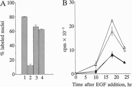
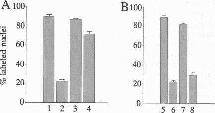
One of the more intriguing aspects of transforming growth factor beta 1 (TGF beta 1) is its ability to function as both a mitogenic factor for certain mesenchymal cells and a potent growth inhibitor of lymphoid, endothelial, and epithelial cells. Data are presented indicating that c-myc may play a pivotal role in both the mitogenic and antiproliferative actions of TGF beta 1. In agreement with previous studies using C3H/10T1/2 fibroblasts constitutively expressing an exogenous c-myc cDNA, we show that AKR-2B fibroblasts expressing a chimeric estrogen-inducible form of c-myc (mycER) are able to form colonies in soft agar in the presence of TGF beta 1 only when c-myc is activated by hormone. Whereas these findings support a synergistic role for c-myc in mitogenic responses to TGF beta 1, we also find that c-myc can antagonize the growth-inhibitory response to TGF beta 1. Mouse keratinocytes (BALB/MK), which are normally growth-arrested by TGF beta 1, are rendered insensitive to the growth-inhibitory effects of TGF beta 1 upon mycER activation. This ability of mycER activation to block TGF beta 1-induced growth arrest was found to occur only when the fusion protein was induced with hormone in the early part of G1. Addition of estradiol late in G1 had no suppressive effect on TGF beta 1-induced growth inhibition.
转化生长因子β1(TGFβ1)较为引人关注的一个方面是,它既能作为某些间充质细胞的促有丝分裂因子,又能作为淋巴细胞、内皮细胞和上皮细胞强大的生长抑制剂。本文提供的数据表明,c-myc可能在TGFβ1的促有丝分裂和抗增殖作用中都发挥关键作用。与之前使用组成性表达外源性c-myc cDNA的C3H/10T1/2成纤维细胞的研究一致,我们发现,表达嵌合雌激素诱导型c-myc(mycER)的AKR-2B成纤维细胞,只有在c-myc被激素激活时,才能在TGFβ1存在的情况下在软琼脂中形成集落。虽然这些发现支持c-myc在对TGFβ1的促有丝分裂反应中起协同作用,但我们也发现c-myc可以拮抗对TGFβ1的生长抑制反应。通常会被TGFβ1生长阻滞的小鼠角质形成细胞(BALB/MK),在mycER激活后对TGFβ1的生长抑制作用变得不敏感。发现只有当在G1期早期用激素诱导融合蛋白时,mycER激活才能阻断TGFβ1诱导的生长阻滞。在G1期后期添加雌二醇对TGFβ1诱导的生长抑制没有抑制作用。