• 文献检索
  • 文档翻译
  • 深度研究
  • 学术资讯
  • Suppr Zotero 插件Zotero 插件
  • 邀请有礼
  • 套餐&价格
  • 历史记录
应用&插件
Suppr Zotero 插件Zotero 插件浏览器插件Mac 客户端Windows 客户端微信小程序
定价
高级版会员购买积分包购买API积分包
服务
文献检索文档翻译深度研究API 文档MCP 服务
关于我们
关于 Suppr公司介绍联系我们用户协议隐私条款
关注我们

Suppr 超能文献

核心技术专利:CN118964589B侵权必究
粤ICP备2023148730 号-1Suppr @ 2026

文献检索

告别复杂PubMed语法,用中文像聊天一样搜索,搜遍4000万医学文献。AI智能推荐,让科研检索更轻松。

立即免费搜索

文件翻译

保留排版,准确专业,支持PDF/Word/PPT等文件格式,支持 12+语言互译。

免费翻译文档

深度研究

AI帮你快速写综述,25分钟生成高质量综述,智能提取关键信息,辅助科研写作。

立即免费体验

通过扫描电子显微镜观察扩张动脉的腔面:消除形态和技术假象

Luminal surface of distended arteries by scanning electron microscopy: eliminating configurational and technical artefacts.

作者信息

Clark J M, Glagov S

出版信息

Br J Exp Pathol. 1976 Feb;57(1):129-35.

PMID:773399
原文链接:https://pmc.ncbi.nlm.nih.gov/articles/PMC2041173/
Abstract

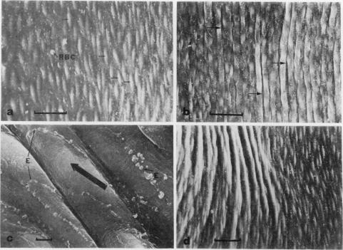
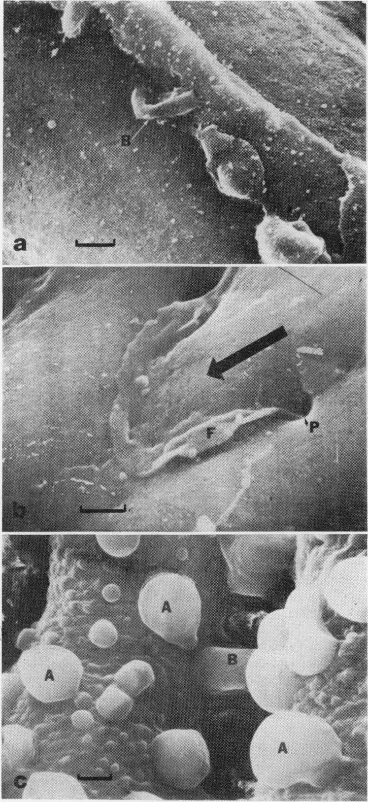
Perfusion fixation at physiological pressures, careful tissue handling, adequate drying and reduced beam exposure time eliminated many of the intimal surface projections, ridges and bridges which have been taken for normal structures on scanning electron microscopy. In the normal distended vessel, ovoid endothelial nuclei bulged into the lumen with their major axes aligned in the direction of flow: adjacent cell margins overlapped consistently in the direction of flow, with each cell overlapping the edge of its downstream neighbour. Regular longitudinal furrows associated with undulations of the internal elastin lamina were entirely eliminated from elastic arteries when distending pressures exceeded diastolic levels during fixation.

摘要

在生理压力下进行灌注固定、小心处理组织、充分干燥并缩短电子束照射时间,消除了许多在扫描电子显微镜下被视为正常结构的内膜表面突起、嵴和桥。在正常扩张的血管中,椭圆形的内皮细胞核向管腔内突出,其长轴与血流方向一致:相邻细胞边缘在血流方向上持续重叠,每个细胞与其下游相邻细胞的边缘重叠。当在固定过程中扩张压力超过舒张压水平时,弹性动脉中与内弹性膜波动相关的规则纵向沟完全消失。

https://cdn.ncbi.nlm.nih.gov/pmc/blobs/23da/2041173/7463e329c8f1/brjexppathol00139-0145-a.jpg
https://cdn.ncbi.nlm.nih.gov/pmc/blobs/23da/2041173/35aaa32c96a8/brjexppathol00139-0143-a.jpg
https://cdn.ncbi.nlm.nih.gov/pmc/blobs/23da/2041173/9dee1889ea22/brjexppathol00139-0144-a.jpg
https://cdn.ncbi.nlm.nih.gov/pmc/blobs/23da/2041173/7463e329c8f1/brjexppathol00139-0145-a.jpg
https://cdn.ncbi.nlm.nih.gov/pmc/blobs/23da/2041173/35aaa32c96a8/brjexppathol00139-0143-a.jpg
https://cdn.ncbi.nlm.nih.gov/pmc/blobs/23da/2041173/9dee1889ea22/brjexppathol00139-0144-a.jpg
https://cdn.ncbi.nlm.nih.gov/pmc/blobs/23da/2041173/7463e329c8f1/brjexppathol00139-0145-a.jpg

相似文献

1
Luminal surface of distended arteries by scanning electron microscopy: eliminating configurational and technical artefacts.通过扫描电子显微镜观察扩张动脉的腔面:消除形态和技术假象
Br J Exp Pathol. 1976 Feb;57(1):129-35.
2
A scanning electron microscope study on the endothelium of the vessels. I. Fine structure of the endothelial surface of aorta and some other arteries in normal rabbits.血管内皮的扫描电子显微镜研究。I. 正常兔主动脉及其他一些动脉内皮表面的精细结构。
Arch Histol Jpn. 1974 Jul;37(1):1-14.
3
Scanning electron microscopic study of the effects of pressure on the luminal surface of the rabbit aorta.压力对兔主动脉管腔表面影响的扫描电子显微镜研究。
Am J Anat. 1976 Jan;145(1):137-42. doi: 10.1002/aja.1001450111.
4
Structure of large arteries: orientation of elastin in rabbit aortic internal elastic lamina and in the elastic lamellae of aortic media.大动脉的结构:兔主动脉内弹性膜及主动脉中膜弹性膜中弹性蛋白的取向
Microvasc Res. 2007 Mar;73(2):95-9. doi: 10.1016/j.mvr.2006.10.005. Epub 2006 Dec 18.
5
Scanning electron microscopy - some methodological considerations.扫描电子显微镜术——一些方法学上的考量
Artery. 1980;8(3):225-35.
6
Morphologic effects of pressure changes on canine carotid artery endothelium as observed by scanning electron microscopy.通过扫描电子显微镜观察压力变化对犬颈动脉内皮的形态学影响。
Anat Rec. 1979 May;194(1):1-14. doi: 10.1002/ar.1091940102.
7
Morphologic investigation of coronary arteries subjected to hypertension by experimental supravalvular aortic stenosis in dogs.犬实验性主动脉瓣上狭窄致高血压状态下冠状动脉的形态学研究
Lab Invest. 1984 Apr;50(4):469-79.
8
Scanning electron microscopic observation of intimal surface of normal and atherosclerotic arteries.正常动脉和动脉粥样硬化动脉内膜表面的扫描电子显微镜观察
Acta Morphol Acad Sci Hung. 1978;26(2):173-84.
9
Repair in arterial tissue. A semiquantitative scanning-electron-microscopic and light-microscopic study on the effects of toxic doses of noradrenaline on the endothelium of the rabbit thoracic aorta.动脉组织的修复。关于去甲肾上腺素中毒剂量对兔胸主动脉内皮影响的半定量扫描电子显微镜和光学显微镜研究。
Blood Vessels. 1977 Mar;14(2):116-27.
10
Arterial elastin as seen with scanning electron microscopy: a review.扫描电子显微镜下观察到的动脉弹性蛋白:综述
Scanning Microsc. 1988 Jun;2(2):994-1004.

引用本文的文献

1
Immediate ultrastructural effects of endothelin-1 on rabbit basilar artery. A comparison between immersion and perfusion fixation techniques.内皮素-1对兔基底动脉的即时超微结构效应。浸泡固定技术与灌注固定技术的比较。
Acta Neurochir (Wien). 1995;132(1-3):104-9. doi: 10.1007/BF01404856.
2
A scanning electron-microscopic analysis of the intraparenchymal brain vasculature following experimental hypertension.实验性高血压后脑实质内血管系统的扫描电子显微镜分析。
Acta Neuropathol. 1980;51(3):203-13. doi: 10.1007/BF00687387.
3
Umbilical vessels of spontaneously hypertensive rats.

本文引用的文献

1
STRUCTURAL BASIS FOR THE STATIC MECHANICAL PROPERTIES OF THE AORTIC MEDIA.主动脉中膜静态力学特性的结构基础
Circ Res. 1964 May;14:400-13. doi: 10.1161/01.res.14.5.400.
2
Endothelial projections as revealed by scanning electron microscopy.扫描电子显微镜显示的内皮细胞突起。
Science. 1971 Sep 3;173(4000):925-7. doi: 10.1126/science.173.4000.925.
3
The arterial endothelium of the hypertensive rat: a scanning and transmission electron microscopical study.高血压大鼠的动脉内皮:扫描电镜与透射电镜研究
Anat Embryol (Berl). 1984;170(3):289-94. doi: 10.1007/BF00318732.
4
Ultrastructure of cerebral arteries following experimental subarachnoid haemorrhage.实验性蛛网膜下腔出血后脑动脉的超微结构
J Neurol Neurosurg Psychiatry. 1985 Mar;48(3):256-62. doi: 10.1136/jnnp.48.3.256.
5
Pathology of the intima in coarctation of the aorta: a study using light and scanning electron microscopy.主动脉缩窄内膜的病理学:一项运用光学显微镜和扫描电子显微镜的研究
Thorax. 1979 Jun;34(3):366-74. doi: 10.1136/thx.34.3.366.
6
Platelet-leukocyte interactions following arterial endothelial injury.动脉内皮损伤后的血小板-白细胞相互作用。
Am J Pathol. 1979 Aug;96(2):567-80.
7
The vascular endothelium-pathobiologic significance.血管内皮——病理生物学意义。
Am J Pathol. 1978 Dec;93(3):803-48.
8
The care of vascular endothelium in pediatric surgery.小儿外科中血管内皮的护理
Ann Surg. 1978 Aug;188(2):216-28. doi: 10.1097/00000658-197808000-00015.
Arch Pathol. 1974 Jun;97(6):337-42.
4
A scanning electron microscopic (SEM) study on the endothelium of the normal rabbit aorta.正常兔主动脉内皮的扫描电子显微镜(SEM)研究。
Angiologica. 1972;9(1):15-26. doi: 10.1159/000157911.
5
Scanning electron microscopy of aortic endothelial cell boundaries after staining with silver nitrate.硝酸银染色后主动脉内皮细胞边界的扫描电子显微镜观察。
Angiologica. 1970;7(6):365-73. doi: 10.1159/000157852.