• 文献检索
  • 文档翻译
  • 深度研究
  • 学术资讯
  • Suppr Zotero 插件Zotero 插件
  • 邀请有礼
  • 套餐&价格
  • 历史记录
应用&插件
Suppr Zotero 插件Zotero 插件浏览器插件Mac 客户端Windows 客户端微信小程序
定价
高级版会员购买积分包购买API积分包
服务
文献检索文档翻译深度研究API 文档MCP 服务
关于我们
关于 Suppr公司介绍联系我们用户协议隐私条款
关注我们

Suppr 超能文献

核心技术专利:CN118964589B侵权必究
粤ICP备2023148730 号-1Suppr @ 2026

文献检索

告别复杂PubMed语法,用中文像聊天一样搜索,搜遍4000万医学文献。AI智能推荐,让科研检索更轻松。

立即免费搜索

文件翻译

保留排版,准确专业,支持PDF/Word/PPT等文件格式,支持 12+语言互译。

免费翻译文档

深度研究

AI帮你快速写综述,25分钟生成高质量综述,智能提取关键信息,辅助科研写作。

立即免费体验

部分性癫痫的磁共振成像:液体衰减反转恢复(FLAIR)脉冲序列显示的额外异常

Magnetic resonance imaging in partial epilepsy: additional abnormalities shown with the fluid attenuated inversion recovery (FLAIR) pulse sequence.

作者信息

Bergin P S, Fish D R, Shorvon S D, Oatridge A, deSouza N M, Bydder G M

机构信息

Institute of Neurology, National Hospital for Neurology and Neurosurgery, Queen Square, London, UK.

出版信息

J Neurol Neurosurg Psychiatry. 1995 Apr;58(4):439-43. doi: 10.1136/jnnp.58.4.439.

DOI:10.1136/jnnp.58.4.439
PMID:7738550
原文链接:https://pmc.ncbi.nlm.nih.gov/articles/PMC1073429/
Abstract

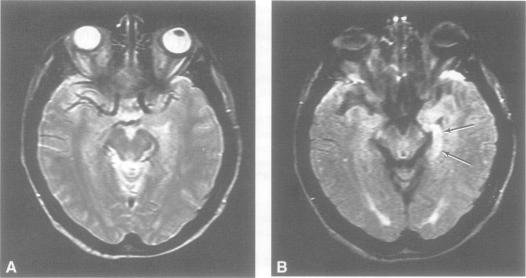
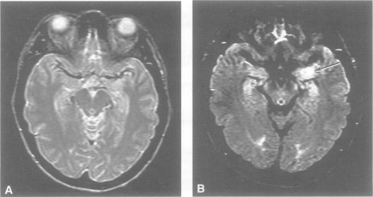
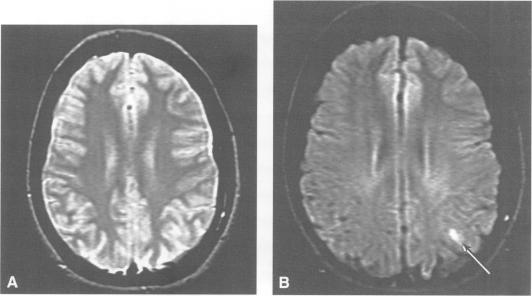
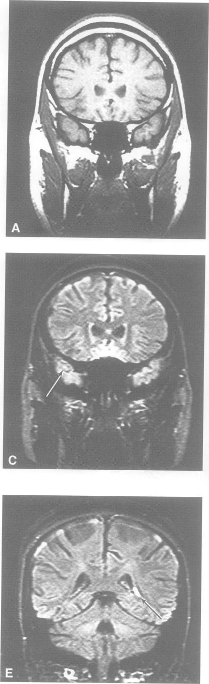
Thirty six patients with a history of partial epilepsy had MRI of the brain performed with conventional T1 and T2 weighted pulse sequences as well as the fluid attenuated inversion recovery (FLAIR) sequence. Abnormalities were found in 20 cases (56%), in whom there were 25 lesions or groups of lesions. Twenty four of these lesions were more conspicuous with the FLAIR sequence than with any of the conventional sequences. In 11 of these 20 cases, lesions thought to be of aetiological importance were only seen with the FLAIR sequence. In eight this was a solitary lesion. In the other three, an additional and apparently significant lesion (or lesions) was only seen with the FLAIR sequence when another lesion had been identified with both conventional and FLAIR sequences. The 11 additional lesions or groups of lesions were seen in the hippocampus, amygdala, cortex, or subcortical and periventricular regions. No lesion was found with any pulse sequence in 16 (44%) of the original group of 36 patients. In the eight cases where a lesion was seen only with the FLAIR sequence, localisation was concordant with the electroclinical features. Two of the eight patients with solitary lesions seen only on the FLAIR sequence underwent surgery, after which there was pathological confirmation of the abnormality identified with imaging. In one patient with a congenital cavernoma, the primary lesion was best seen with a contrast enhanced T1 weighted spin echo sequence. In this selected series, the FLAIR sequence increased the yield of MRI examinations of the brain by 30%.

摘要

36例有部分性癫痫病史的患者接受了脑部MRI检查,采用传统的T1加权和T2加权脉冲序列以及液体衰减反转恢复(FLAIR)序列。20例(56%)发现异常,其中有25个病灶或病灶群。这些病灶中有24个在FLAIR序列上比在任何传统序列上更明显。在这20例中的11例中,被认为具有病因学重要性的病灶仅在FLAIR序列上可见。其中8例为单个病灶。在另外3例中,当另一个病灶在传统序列和FLAIR序列上均被识别出来时,一个额外的且明显有意义的病灶(或多个病灶)仅在FLAIR序列上可见。这11个额外的病灶或病灶群见于海马体、杏仁核、皮质或皮质下及脑室周围区域。在最初的36例患者中,16例(44%)在任何脉冲序列上均未发现病灶。在仅在FLAIR序列上发现病灶的8例中,病灶定位与电临床特征一致。仅在FLAIR序列上发现单个病灶的8例患者中有2例接受了手术,术后病理证实了影像学上发现的异常。在1例先天性海绵状血管瘤患者中,主要病灶在对比增强T1加权自旋回波序列上显示最佳。在这个选定的系列中,FLAIR序列使脑部MRI检查的检出率提高了30%。

https://cdn.ncbi.nlm.nih.gov/pmc/blobs/50d9/1073429/608d4a081f87/jnnpsyc00028-0046-b.jpg
https://cdn.ncbi.nlm.nih.gov/pmc/blobs/50d9/1073429/32305b6014d3/jnnpsyc00028-0044-a.jpg
https://cdn.ncbi.nlm.nih.gov/pmc/blobs/50d9/1073429/caf5b2f727d4/jnnpsyc00028-0044-b.jpg
https://cdn.ncbi.nlm.nih.gov/pmc/blobs/50d9/1073429/d5bdeb628b56/jnnpsyc00028-0045-a.jpg
https://cdn.ncbi.nlm.nih.gov/pmc/blobs/50d9/1073429/54423d12a4cc/jnnpsyc00028-0045-b.jpg
https://cdn.ncbi.nlm.nih.gov/pmc/blobs/50d9/1073429/490d9cbadc41/jnnpsyc00028-0046-a.jpg
https://cdn.ncbi.nlm.nih.gov/pmc/blobs/50d9/1073429/608d4a081f87/jnnpsyc00028-0046-b.jpg

相似文献

1
Magnetic resonance imaging in partial epilepsy: additional abnormalities shown with the fluid attenuated inversion recovery (FLAIR) pulse sequence.部分性癫痫的磁共振成像:液体衰减反转恢复(FLAIR)脉冲序列显示的额外异常
J Neurol Neurosurg Psychiatry. 1995 Apr;58(4):439-43. doi: 10.1136/jnnp.58.4.439.
2
Yield of fluid-attenuated inversion recovery in drug-resistant focal epilepsy with noninformative conventional magnetic resonance imaging.在传统磁共振成像无信息的耐药性局灶性癫痫中,液体衰减反转恢复序列的检出率。
Eur Neurol. 1999;41(2):64-72. doi: 10.1159/000008005.
3
T1-weighted fluid-attenuated inversion recovery and T1-weighted fast spin-echo contrast-enhanced imaging: a comparison in 20 patients with brain lesions.T1加权液体衰减反转恢复序列与T1加权快速自旋回波对比增强成像:20例脑病变患者的比较
J Med Imaging Radiat Oncol. 2009 Aug;53(4):366-72. doi: 10.1111/j.1754-9485.2009.02093.x.
4
Comparing lesion detection of infratentorial multiple sclerosis lesions between T2-weighted spin-echo, 2D-FLAIR, and 3D-FLAIR sequences.比较T2加权自旋回波序列、二维液体衰减反转恢复序列(2D-FLAIR)和三维液体衰减反转恢复序列(3D-FLAIR)对幕下多发性硬化病变的检测情况。
Clin Imaging. 2018 Sep-Oct;51:229-234. doi: 10.1016/j.clinimag.2018.05.017. Epub 2018 May 29.
5
Usefulness of optimized gadolinium-enhanced fast fluid-attenuated inversion recovery MR imaging in revealing lesions of the brain.优化的钆增强快速液体衰减反转恢复磁共振成像在显示脑病变中的效用。
AJR Am J Roentgenol. 1998 Sep;171(3):803-7. doi: 10.2214/ajr.171.3.9725320.
6
Diagnostic value of 3D fluid attenuated inversion recovery sequence in multiple sclerosis.三维液体衰减反转恢复序列在多发性硬化症中的诊断价值
Acta Radiol. 2015 May;56(5):622-7. doi: 10.1177/0284185114534413. Epub 2014 May 27.
7
Relationship between contrast enhancement on fluid-attenuated inversion recovery MR sequences and signal intensity on T2-weighted MR images: visual evaluation of brain tumors.液体衰减反转恢复磁共振序列上的对比增强与T2加权磁共振图像上的信号强度之间的关系:脑肿瘤的视觉评估
J Magn Reson Imaging. 2005 Jun;21(6):694-700. doi: 10.1002/jmri.20331.
8
Diagnostic value of contrast-enhanced fluid-attenuated inversion-recovery MRI for intracranial tumors in comparison with post-contrast T1W spin-echo MRI.与对比增强T1加权自旋回波MRI相比,对比增强液体衰减反转恢复序列MRI对颅内肿瘤的诊断价值
Chin Med J (Engl). 2006 Mar 20;119(6):467-73.
9
Neuroradiological findings in patients with "non-lesional" focal epilepsy revealed by research protocol.根据研究方案,在“非病灶性”局灶性癫痫患者中发现的神经放射学表现。
Clin Radiol. 2019 Jan;74(1):78.e1-78.e11. doi: 10.1016/j.crad.2018.08.013. Epub 2018 Sep 28.
10
Comparison of contrast-enhanced T1-weighted FLAIR with BLADE, and spin-echo T1-weighted sequences in intracranial MRI.颅内磁共振成像中对比增强T1加权液体衰减反转恢复序列与刀锋序列及自旋回波T1加权序列的比较。
Diagn Interv Radiol. 2009 Jun;15(2):75-80.

引用本文的文献

1
A brain tumor computer-aided diagnosis method with automatic lesion segmentation and ensemble decision strategy.一种具有自动病变分割和集成决策策略的脑肿瘤计算机辅助诊断方法。
Front Med (Lausanne). 2023 Sep 29;10:1232496. doi: 10.3389/fmed.2023.1232496. eCollection 2023.
2
Morphological and Advanced Imaging of Epilepsy: Beyond the Basics.癫痫的形态学与高级影像学:基础之外
Children (Basel). 2019 Mar 11;6(3):43. doi: 10.3390/children6030043.
3
The value of repeat neuroimaging for epilepsy at a tertiary referral centre: 16 years of experience.

本文引用的文献

1
Detection of hippocampal pathology in intractable partial epilepsy: increased sensitivity with quantitative magnetic resonance T2 relaxometry.难治性部分性癫痫中海马病变的检测:定量磁共振T2弛豫测量法提高了敏感性。
Neurology. 1993 Sep;43(9):1793-9. doi: 10.1212/wnl.43.9.1793.
2
RARE imaging: a fast imaging method for clinical MR.RARE成像:一种用于临床磁共振成像的快速成像方法。
Magn Reson Med. 1986 Dec;3(6):823-33. doi: 10.1002/mrm.1910030602.
3
Complex partial seizures and small posterior temporal or extratemporal structural lesions: surgical management.
三级转诊中心癫痫重复神经影像学的价值:16 年的经验。
Epilepsy Res. 2013 Aug;105(3):349-55. doi: 10.1016/j.eplepsyres.2013.02.022. Epub 2013 Mar 26.
4
Presurgical evaluation and surgical treatment of medically refractory epilepsy.药物难治性癫痫的术前评估与手术治疗
Neurosurg Rev. 2004 Jan;27(1):1-18; discussion 19-21. doi: 10.1007/s10143-003-0305-6. Epub 2003 Oct 28.
5
Clinical application of neuroimaging in epilepsy.神经影像学在癫痫中的临床应用。
J Neurol Neurosurg Psychiatry. 2003 Apr;74(4):466-70. doi: 10.1136/jnnp.74.4.466.
6
Transient MR signal changes in patients with generalized tonicoclonic seizure or status epilepticus: periictal diffusion-weighted imaging.全身强直阵挛性发作或癫痫持续状态患者的短暂磁共振信号变化:发作期弥散加权成像
AJNR Am J Neuroradiol. 2001 Jun-Jul;22(6):1149-60.
7
Applicability and advantages of flow artifact-insensitive fluid-attenuated inversion-recovery MR sequences for imaging the posterior fossa.对后颅窝成像的血流伪影不敏感的液体衰减反转恢复磁共振序列的适用性及优势
AJNR Am J Neuroradiol. 2000 Jun-Jul;21(6):1095-8.
8
Magnetic resonance imaging in epilepsy with a fast FLAIR sequence.采用快速液体衰减反转恢复序列对癫痫进行磁共振成像
J Neurol Neurosurg Psychiatry. 1996 Oct;61(4):357-61. doi: 10.1136/jnnp.61.4.357.
9
High resolution magnetic resonance imaging in adults with partial or secondary generalised epilepsy attending a tertiary referral unit.在一家三级转诊单位就诊的部分性或继发性全身性癫痫成年患者中的高分辨率磁共振成像。
J Neurol Neurosurg Psychiatry. 1995 Oct;59(4):384-7. doi: 10.1136/jnnp.59.4.384.
复杂部分性发作与颞叶后部或颞叶外小结构病变:手术治疗
Neurology. 1991 Nov;41(11):1781-4. doi: 10.1212/wnl.41.11.1781.
4
MR of the brain using fluid-attenuated inversion recovery (FLAIR) pulse sequences.使用液体衰减反转恢复(FLAIR)脉冲序列的脑部磁共振成像。
AJNR Am J Neuroradiol. 1992 Nov-Dec;13(6):1555-64.