• 文献检索
  • 文档翻译
  • 深度研究
  • 学术资讯
  • Suppr Zotero 插件Zotero 插件
  • 邀请有礼
  • 套餐&价格
  • 历史记录
应用&插件
Suppr Zotero 插件Zotero 插件浏览器插件Mac 客户端Windows 客户端微信小程序
定价
高级版会员购买积分包购买API积分包
服务
文献检索文档翻译深度研究API 文档MCP 服务
关于我们
关于 Suppr公司介绍联系我们用户协议隐私条款
关注我们

Suppr 超能文献

核心技术专利:CN118964589B侵权必究
粤ICP备2023148730 号-1Suppr @ 2026

文献检索

告别复杂PubMed语法,用中文像聊天一样搜索,搜遍4000万医学文献。AI智能推荐,让科研检索更轻松。

立即免费搜索

文件翻译

保留排版,准确专业,支持PDF/Word/PPT等文件格式,支持 12+语言互译。

免费翻译文档

深度研究

AI帮你快速写综述,25分钟生成高质量综述,智能提取关键信息,辅助科研写作。

立即免费体验

增殖细胞核抗原、波形蛋白、c-Fos和簇集蛋白在缺血后肾脏中的定位。肾单位节段间存在异质性基因反应以及大量有丝分裂活跃和去分化细胞的证据。

Localization of proliferating cell nuclear antigen, vimentin, c-Fos, and clusterin in the postischemic kidney. Evidence for a heterogenous genetic response among nephron segments, and a large pool of mitotically active and dedifferentiated cells.

作者信息

Witzgall R, Brown D, Schwarz C, Bonventre J V

机构信息

Renal Unit, Medical Services, Massachusetts General Hospital East, Charlestown 02129.

出版信息

J Clin Invest. 1994 May;93(5):2175-88. doi: 10.1172/JCI117214.

DOI:10.1172/JCI117214
PMID:7910173
原文链接:https://pmc.ncbi.nlm.nih.gov/articles/PMC294357/
Abstract

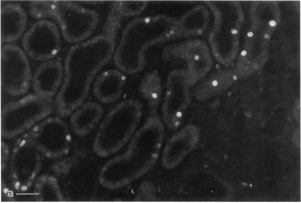
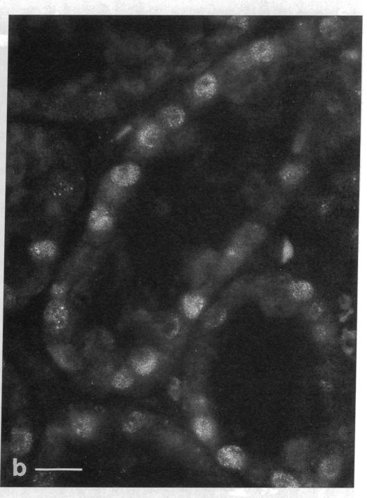
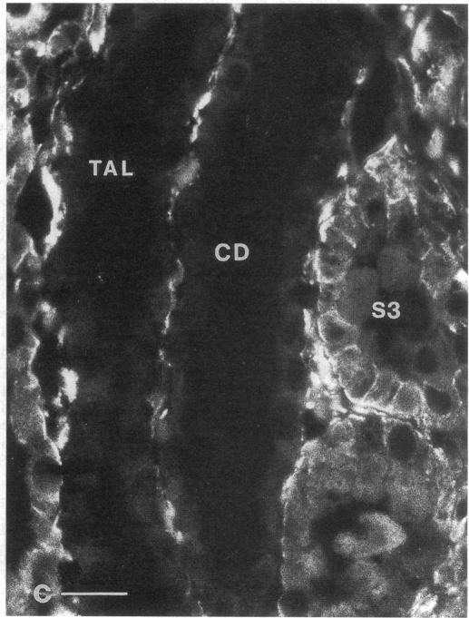
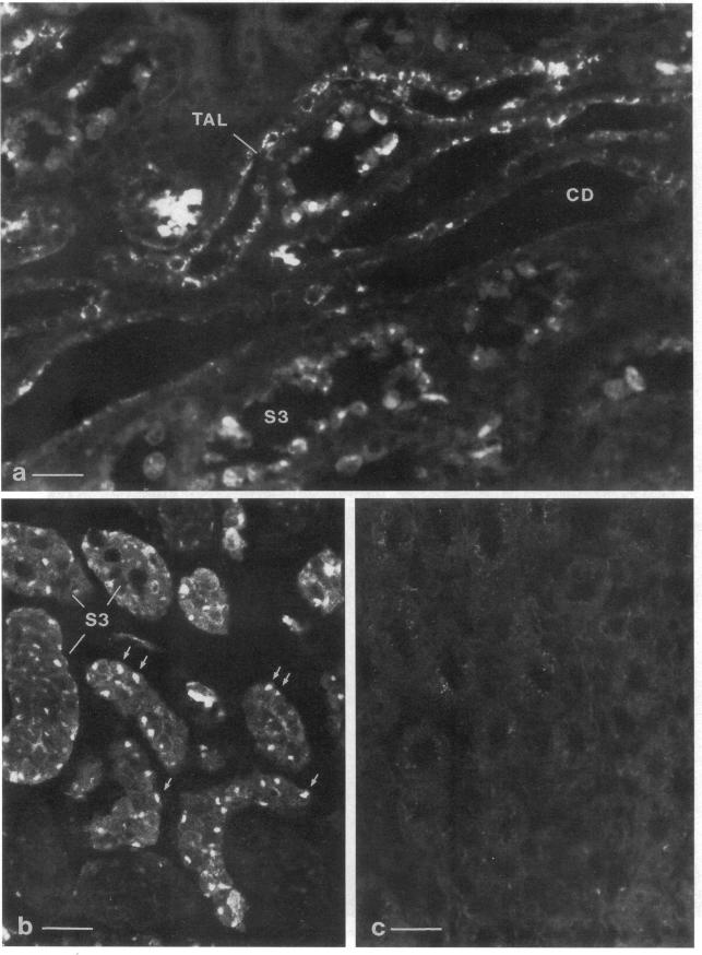
The mechanisms leading to the recovery of the kidney after ischemic acute renal failure are poorly understood. To explore the role played by mitogenesis and dedifferentiation in this repair process and to identify whether the genetic response of the nephron segments reflects the level of susceptibility to injury, the temporal and nephron segment expressions of various proteins implicated in mitogenesis, differentiation, and injury were determined. Proliferating cell nuclear antigen (PCNA), a marker for the G1-S transition in the cell cycle and hence mitogenesis, was detected primarily in the S3 segment of the proximal tubule, with maximal expression at 2 d postischemia. Vimentin, normally present in mesenchymal cells but not epithelial cells, and hence a marker for the state of differentiation, was prominently expressed in the S3 segment 2-5 d postischemia. In the S3 segments in the outer stripe of the medulla cells that stained positively for PCNA also stained positively for vimentin. Clusterin, a marker for cell injury, was expressed primarily in the S3 segment and in the distal tubule with distinct staining patterns in each segment. None of the cells that stained with clusterin antibodies were positively stained with PCNA or vimentin antibodies. Likewise, none of the PCNA or vimentin-positive cells expressed clusterin at detectable levels. Thus, in the S3 segment, where there is significant ischemic injury, surviving cells express markers indicating that they undergo mitogenesis and dedifferentiate in the postischemic period. While there is some expression of c-Fos in the S3 segment, c-Fos was expressed predominantly, at 1 and 3 h postischemia, in the nuclei of the distal nephron, particularly in the thick ascending limb. The data support the view that the mature renal S3 segment epithelial cell can be a progenitor cell.

摘要

缺血性急性肾衰竭后肾脏恢复的机制尚不清楚。为了探究有丝分裂和去分化在这一修复过程中所起的作用,并确定肾单位各节段的基因反应是否反映了对损伤的易感性水平,我们测定了参与有丝分裂、分化和损伤的各种蛋白质的时间和肾单位节段表达情况。增殖细胞核抗原(PCNA)是细胞周期中G1-S期转换的标志物,因此也是有丝分裂的标志物,主要在近端小管的S3节段中检测到,在缺血后2天表达量最高。波形蛋白通常存在于间充质细胞而非上皮细胞中,因此是分化状态的标志物,在缺血后2-5天在S3节段中显著表达。在髓质外带的S3节段中,对PCNA染色呈阳性的细胞对波形蛋白染色也呈阳性。聚集素是细胞损伤的标志物,主要在S3节段和远端小管中表达,且在每个节段中有不同的染色模式。用聚集素抗体染色的细胞均未被PCNA或波形蛋白抗体阳性染色。同样,PCNA或波形蛋白阳性的细胞也未检测到可检测水平的聚集素表达。因此,在存在严重缺血损伤的S3节段中,存活的细胞表达的标志物表明它们在缺血后期经历了有丝分裂和去分化。虽然S3节段中有一些c-Fos表达,但c-Fos在缺血后1小时和3小时主要在远端肾单位的细胞核中表达,特别是在厚壁升支中。这些数据支持成熟的肾S3节段上皮细胞可以是祖细胞的观点。

https://cdn.ncbi.nlm.nih.gov/pmc/blobs/b142/294357/89ee2a45a66f/jcinvest00034-0323-a.jpg
https://cdn.ncbi.nlm.nih.gov/pmc/blobs/b142/294357/ac8a4bce1f94/jcinvest00034-0312-a.jpg
https://cdn.ncbi.nlm.nih.gov/pmc/blobs/b142/294357/0f1606e6f74d/jcinvest00034-0312-b.jpg
https://cdn.ncbi.nlm.nih.gov/pmc/blobs/b142/294357/ebd5761e9f81/jcinvest00034-0313-a.jpg
https://cdn.ncbi.nlm.nih.gov/pmc/blobs/b142/294357/adaf35c648ae/jcinvest00034-0313-b.jpg
https://cdn.ncbi.nlm.nih.gov/pmc/blobs/b142/294357/85326e62f2dd/jcinvest00034-0314-a.jpg
https://cdn.ncbi.nlm.nih.gov/pmc/blobs/b142/294357/549571b38e6f/jcinvest00034-0314-b.jpg
https://cdn.ncbi.nlm.nih.gov/pmc/blobs/b142/294357/175ae5494c05/jcinvest00034-0316-a.jpg
https://cdn.ncbi.nlm.nih.gov/pmc/blobs/b142/294357/04a006c51f18/jcinvest00034-0316-b.jpg
https://cdn.ncbi.nlm.nih.gov/pmc/blobs/b142/294357/5ce0b87cd0f8/jcinvest00034-0316-c.jpg
https://cdn.ncbi.nlm.nih.gov/pmc/blobs/b142/294357/d990aa6d3df3/jcinvest00034-0317-a.jpg
https://cdn.ncbi.nlm.nih.gov/pmc/blobs/b142/294357/ca2b58e1312f/jcinvest00034-0318-a.jpg
https://cdn.ncbi.nlm.nih.gov/pmc/blobs/b142/294357/cb772371d40c/jcinvest00034-0318-b.jpg
https://cdn.ncbi.nlm.nih.gov/pmc/blobs/b142/294357/d6b76a90ed33/jcinvest00034-0319-a.jpg
https://cdn.ncbi.nlm.nih.gov/pmc/blobs/b142/294357/e528d4cf3bd9/jcinvest00034-0320-a.jpg
https://cdn.ncbi.nlm.nih.gov/pmc/blobs/b142/294357/9b60a94b57a2/jcinvest00034-0322-a.jpg
https://cdn.ncbi.nlm.nih.gov/pmc/blobs/b142/294357/89ee2a45a66f/jcinvest00034-0323-a.jpg

相似文献

1
Localization of proliferating cell nuclear antigen, vimentin, c-Fos, and clusterin in the postischemic kidney. Evidence for a heterogenous genetic response among nephron segments, and a large pool of mitotically active and dedifferentiated cells.增殖细胞核抗原、波形蛋白、c-Fos和簇集蛋白在缺血后肾脏中的定位。肾单位节段间存在异质性基因反应以及大量有丝分裂活跃和去分化细胞的证据。
J Clin Invest. 1994 May;93(5):2175-88. doi: 10.1172/JCI117214.
2
Expression of NCAM recapitulates tubulogenic development in kidneys recovering from acute ischemia.NCAM的表达概括了急性缺血后恢复中的肾脏的肾小管生成发育过程。
Am J Physiol. 1999 Sep;277(3):F454-63. doi: 10.1152/ajprenal.1999.277.3.F454.
3
Cell division and phenotypic regression of proximal tubular cells in response to uranyl acetate insult in rats.大鼠近端肾小管细胞对醋酸铀损伤的细胞分裂及表型消退
Nephrol Dial Transplant. 2009 Sep;24(9):2686-92. doi: 10.1093/ndt/gfp199. Epub 2009 Apr 25.
4
Hyperplasia, hypertrophy, and phenotypic alterations in the distal nephron after acute proximal tubular injury in the rat.大鼠急性近端肾小管损伤后远端肾单位的增生、肥大及表型改变
Lab Invest. 1994 Apr;70(4):479-93.
5
Obstructive nephropathy in the neonatal rat is attenuated by epidermal growth factor.表皮生长因子可减轻新生大鼠的梗阻性肾病。
Kidney Int. 1998 Jul;54(1):38-47. doi: 10.1046/j.1523-1755.1998.00966.x.
6
Clusterin expression and apoptosis in tissue remodeling associated with renal regeneration.与肾再生相关的组织重塑中的簇集蛋白表达与细胞凋亡。
Kidney Int. 1995 Feb;47(2):411-20. doi: 10.1038/ki.1995.54.
7
The type I iodothyronine 5'-deiodinase messenger ribonucleic acid is localized to the S3 segment of the rat kidney proximal tubule.I型碘甲腺原氨酸5'-脱碘酶信使核糖核酸定位于大鼠肾近端小管的S3段。
Endocrinology. 1993 May;132(5):2136-40. doi: 10.1210/endo.132.5.8477661.
8
Expression of Id-1 mRNA and protein in the post-ischemic regenerating rat kidney.
Exp Nephrol. 1998 May-Jun;6(3):253-64. doi: 10.1159/000020530.
9
Differential regulation of B/K protein expression in proximal and distal tubules of rat kidneys with ischemia-reperfusion injury.缺血再灌注损伤大鼠肾脏近端和远端小管中B/K蛋白表达的差异调节
Am J Physiol Renal Physiol. 2007 Jan;292(1):F100-6. doi: 10.1152/ajprenal.00009.2006. Epub 2006 Aug 8.
10
Renal tissue expression of EGF and EGF receptor after ischaemic tubular injury: an immunohistochemical study.缺血性肾小管损伤后表皮生长因子(EGF)及EGF受体的肾组织表达:一项免疫组织化学研究
Exp Nephrol. 1994 Jul-Aug;2(4):229-39.

引用本文的文献

1
Meprin β activity modulates cellular proliferation via trans-signaling IL-6-mediated AKT/ERK pathway in IR-induced kidney injury.在辐射诱导的肾损伤中,金属蛋白酶解整合素与金属蛋白酶-β(Meprin β)活性通过反式信号传导白介素-6(IL-6)介导的AKT/ERK途径调节细胞增殖。
BMC Nephrol. 2025 Aug 18;26(1):467. doi: 10.1186/s12882-025-04224-x.
2
Transplantation of human kidney organoids elicited a robust allogeneic response in a humanized mouse model.在人源化小鼠模型中,人肾类器官移植引发了强烈的同种异体反应。
Kidney Int. 2025 Jun;107(6):1011-1016. doi: 10.1016/j.kint.2025.02.027. Epub 2025 Mar 22.
3
Meprin β activity modulates cellular proliferation via trans-signaling IL-6-mediated AKT/ERK pathway in IR-induced kidney injury.

本文引用的文献

1
Mechanisms of ischemic acute renal failure.缺血性急性肾衰竭的机制。
Kidney Int. 1993 May;43(5):1160-78. doi: 10.1038/ki.1993.163.
2
Kid-1, a putative renal transcription factor: regulation during ontogeny and in response to ischemia and toxic injury.Kid-1,一种假定的肾脏转录因子:个体发育过程中的调控以及对缺血和毒性损伤的反应。
Mol Cell Biol. 1993 Mar;13(3):1933-42. doi: 10.1128/mcb.13.3.1933-1942.1993.
3
Antibody to intercellular adhesion molecule 1 protects the kidney against ischemic injury.细胞间黏附分子1抗体可保护肾脏免受缺血性损伤。
金属蛋白酶β活性通过跨信号传导白介素-6介导的AKT/ERK途径调节辐射诱导的肾损伤中的细胞增殖。
Res Sq. 2025 Jan 30:rs.3.rs-5901359. doi: 10.21203/rs.3.rs-5901359/v1.
4
The Kidney Precision Medicine Project and Single-Cell Biology of the Injured Proximal Tubule.肾脏精准医学项目与近端小管损伤的单细胞生物学
Am J Pathol. 2025 Jan;195(1):7-22. doi: 10.1016/j.ajpath.2024.09.006. Epub 2024 Sep 26.
5
Podocyte-specific KLF6 primes proximal tubule CaMK1D signaling to attenuate diabetic kidney disease.足细胞特异性 KLF6 激活近端肾小管 CaMK1D 信号通路,从而减轻糖尿病肾病。
Nat Commun. 2024 Sep 13;15(1):8038. doi: 10.1038/s41467-024-52306-5.
6
Spatial transcriptomics defines injury specific microenvironments and cellular interactions in kidney regeneration and disease.空间转录组学定义了肾脏再生和疾病中损伤特异性的微环境和细胞相互作用。
Nat Commun. 2024 Sep 5;15(1):7010. doi: 10.1038/s41467-024-51186-z.
7
Injury-induced Foxm1 expression in the mouse kidney drives epithelial proliferation by a cyclin F-dependent mechanism.损伤诱导的小鼠肾脏中Foxm1表达通过细胞周期蛋白F依赖性机制驱动上皮细胞增殖。
JCI Insight. 2024 Jun 25;9(15):e175416. doi: 10.1172/jci.insight.175416.
8
Mesenchymal Stromal Cells Nanovesicles Carry microRNA with Nephroprotective Proprieties Regardless of Aging.间质基质细胞纳米囊泡携带具有肾保护特性的 microRNA,与衰老无关。
Curr Aging Sci. 2024;17(2):118-126. doi: 10.2174/0118746098272926231107061047.
9
Cellular dynamics in pig-to-human kidney xenotransplantation.猪到人肾脏异种移植中的细胞动力学。
Med. 2024 Aug 9;5(8):1016-1029.e4. doi: 10.1016/j.medj.2024.05.003. Epub 2024 May 21.
10
Single-cell transcriptomic analysis reveals transcript enrichment in oxidative phosphorylation, fluid sheer stress, and inflammatory pathways in obesity-related glomerulopathy.单细胞转录组分析揭示了肥胖相关肾小球病中氧化磷酸化、流体切应力和炎症通路中的转录本富集。
Genes Dis. 2023 Sep 14;11(4):101101. doi: 10.1016/j.gendis.2023.101101. eCollection 2024 Jul.
Proc Natl Acad Sci U S A. 1994 Jan 18;91(2):812-6. doi: 10.1073/pnas.91.2.812.
4
Evaluation of proliferating cell nuclear antigen (PCNA) as an endogenous marker of cell proliferation in rat liver: a dual-stain comparison with 5-bromo-2'-deoxyuridine.增殖细胞核抗原(PCNA)作为大鼠肝脏细胞增殖内源性标志物的评估:与5-溴-2'-脱氧尿苷的双重染色比较
J Histochem Cytochem. 1993 Jan;41(1):1-6. doi: 10.1177/41.1.7678022.
5
Effects of verapamil in models of ischemic acute renal failure in the rat.维拉帕米对大鼠缺血性急性肾衰竭模型的影响。
Am J Physiol. 1983 Dec;245(6):F735-42. doi: 10.1152/ajprenal.1983.245.6.F735.
6
Monoclonal antibodies specific for vimentin.针对波形蛋白的单克隆抗体。
Eur J Cell Biol. 1984 May;34(1):137-43.
7
[Ultrastructural detection of the heterogeneity of glycocalyx in convoluted and straight proximal tubules of rat kidney by the lectin-gold complex technic].
C R Seances Acad Sci III. 1983;297(10):501-6.
8
Expression of vimentin and cytokeratin types of intermediate filament proteins in developing and adult human kidneys.波形蛋白和细胞角蛋白类型的中间丝蛋白在发育中和成人肾脏中的表达。
Lab Invest. 1984 May;50(5):552-9.
9
Inhibition of cellular proliferation by antisense oligodeoxynucleotides to PCNA cyclin.通过针对增殖细胞核抗原细胞周期蛋白的反义寡脱氧核苷酸抑制细胞增殖。
Science. 1988 Jun 10;240(4858):1544-6. doi: 10.1126/science.2897717.
10
Coordinated leading and lagging strand synthesis during SV40 DNA replication in vitro requires PCNA.在体外进行SV40 DNA复制过程中,前导链与后随链的协同合成需要增殖细胞核抗原(PCNA)。
Cell. 1988 Apr 8;53(1):117-26. doi: 10.1016/0092-8674(88)90493-x.