Bani D, Riva A, Bigazzi M, Bani Sacchi T
Department of Human Anatomy and Histology, University of Florence, Italy.
Br J Cancer. 1994 Nov;70(5):900-4. doi: 10.1038/bjc.1994.417.
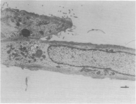
Our previous studies showed that relaxin promotes differentiation of MCF-7 breast adenocarcinoma cells. In the current investigation, we aimed to elucidate whether the effect of the hormone is potentiated when MCF-7 cells are grown together with myoepithelial cells, thus creating a microenvironment reminiscent of the organised tissue architecture of the mammary parenchyma in vivo. The findings obtained reveal that most MCF-7 cells cultured alone have an undifferentiated, blast-like phenotype, only a minority showing a more differentiated phenotype with more organelles and rudimentary intercellular junctions. When co-cultured with myoepithelial cells more MCF-7 cells acquire ultrastructural features consistent with a more differentiated phenotype, such as a rich organellular complement, apical microvilli and intercellular junctions. When relaxin was added to the co-cultures, the ultrastructural signs of differentiation could be observed in even more MCF-7 cells and became more pronounced than in the absence of the hormone, judged by the appearance of a clear-cut polarisation of cytoplasmic organelles, an almost continuous coat of apical microvilli and numerous intracellular pseudolumina.
我们之前的研究表明,松弛素可促进MCF-7乳腺腺癌细胞的分化。在当前的研究中,我们旨在阐明当MCF-7细胞与肌上皮细胞共同培养时,该激素的作用是否会增强,从而形成一种类似于体内乳腺实质有组织的组织结构的微环境。所获得的研究结果显示,大多数单独培养的MCF-7细胞具有未分化的、胚样表型,只有少数细胞表现出更分化的表型,具有更多细胞器和原始的细胞间连接。当与肌上皮细胞共培养时,更多的MCF-7细胞获得了与更分化表型一致的超微结构特征,如丰富的细胞器、顶端微绒毛和细胞间连接。当向共培养物中添加松弛素时,在更多的MCF-7细胞中可观察到分化的超微结构迹象,并且与未添加该激素时相比变得更加明显,这可通过细胞质细胞器的明显极化、几乎连续的顶端微绒毛覆盖以及大量细胞内假腔的出现来判断。