• 文献检索
  • 文档翻译
  • 深度研究
  • 学术资讯
  • Suppr Zotero 插件Zotero 插件
  • 邀请有礼
  • 套餐&价格
  • 历史记录
应用&插件
Suppr Zotero 插件Zotero 插件浏览器插件Mac 客户端Windows 客户端微信小程序
定价
高级版会员购买积分包购买API积分包
服务
文献检索文档翻译深度研究API 文档MCP 服务
关于我们
关于 Suppr公司介绍联系我们用户协议隐私条款
关注我们

Suppr 超能文献

核心技术专利:CN118964589B侵权必究
粤ICP备2023148730 号-1Suppr @ 2026

文献检索

告别复杂PubMed语法,用中文像聊天一样搜索,搜遍4000万医学文献。AI智能推荐,让科研检索更轻松。

立即免费搜索

文件翻译

保留排版,准确专业,支持PDF/Word/PPT等文件格式,支持 12+语言互译。

免费翻译文档

深度研究

AI帮你快速写综述,25分钟生成高质量综述,智能提取关键信息,辅助科研写作。

立即免费体验

C-erbB-2免疫染色:解读问题

C-erbB-2 immunostaining: problems with interpretation.

作者信息

Kay E W, Walsh C J, Cassidy M, Curran B, Leader M

机构信息

Department of Pathology, Royal College of Surgeons, Ireland.

出版信息

J Clin Pathol. 1994 Sep;47(9):816-22. doi: 10.1136/jcp.47.9.816.

DOI:10.1136/jcp.47.9.816
PMID:7962650
原文链接:https://pmc.ncbi.nlm.nih.gov/articles/PMC494938/
Abstract

AIMS

To assess the consistency and reproducibility of assessment of c-erbB-2 immunostaining, and to examine some of the problems relating to inter- and intraobserver variability in the documentation of positive staining; to profile the spectrum of cytoplasmic and membranous staining in a wide range of tumour types.

METHODS

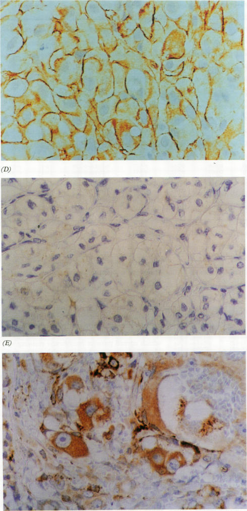
A total of 283 neoplasms were examined for immunohistochemical expression of the c-erbB-2 oncoprotein. Three independent observers were required to assess intensity both of membrane and cytoplasmic staining on a three point and then a four point scale. Extent of positive staining was also assessed on a two point scale. A minimum of two weeks elapsed between assessments using the differing scales.

RESULTS

Positive membrane staining was documented by one or more observers in 16.6% of tumours examined. This positivity was largely restricted to bladder, renal, and breast tumours. The overall level of disagreement as to the presence or absence of membranous staining was 11.3%. Cytoplasmic staining was identified in 55.5% of tumours studied. The level of disagreement as to the presence or absence of cytoplasmic staining was 26.5%.

CONCLUSIONS

Intraobserver variability was minimal, indicating that each pathologist was adhering to internal reproducible standards. Interobserver variability was greater, indicating that the interpretation of c-erbB-2 immunostaining may require set guidelines. It is suggested that assessment should be referenced to a standard positive control, that a three tier system for grading of intensity and a two tier system for grading of extent should be adopted, and that the evaluation should be agreed by at least two pathologists. The presence of cytoplasmic staining should continue to be routinely recorded until its biological role and clinical implications are fully understood.

摘要

目的

评估c-erbB-2免疫染色评估的一致性和可重复性,并研究阳性染色记录中与观察者间和观察者内变异性相关的一些问题;描绘多种肿瘤类型中细胞质和细胞膜染色的谱系。

方法

共检查了283个肿瘤的c-erbB-2癌蛋白的免疫组化表达。要求三名独立观察者在三点然后四点量表上评估细胞膜和细胞质染色的强度。阳性染色范围也在两点量表上进行评估。使用不同量表进行评估之间至少间隔两周。

结果

在检查的肿瘤中,16.6%的肿瘤有一名或多名观察者记录到阳性细胞膜染色。这种阳性主要局限于膀胱、肾脏和乳腺肿瘤。关于细胞膜染色是否存在的总体分歧水平为11.3%。在研究的肿瘤中,55.5%发现有细胞质染色。关于细胞质染色是否存在的分歧水平为26.5%。

结论

观察者内变异性最小,表明每位病理学家都遵循内部可重复的标准。观察者间变异性更大,表明c-erbB-2免疫染色的解释可能需要设定指导原则。建议评估应参考标准阳性对照,采用强度分级的三级系统和范围分级的二级系统,并且评估应由至少两名病理学家达成一致。在细胞质染色的生物学作用和临床意义被充分理解之前,应继续常规记录其存在情况。

https://cdn.ncbi.nlm.nih.gov/pmc/blobs/1b12/494938/3768ff3a5fb3/jclinpath00222-0047-b.jpg
https://cdn.ncbi.nlm.nih.gov/pmc/blobs/1b12/494938/0142ddc31cc4/jclinpath00222-0046-a.jpg
https://cdn.ncbi.nlm.nih.gov/pmc/blobs/1b12/494938/672763936ad9/jclinpath00222-0046-b.jpg
https://cdn.ncbi.nlm.nih.gov/pmc/blobs/1b12/494938/c2360218d4f3/jclinpath00222-0047-a.jpg
https://cdn.ncbi.nlm.nih.gov/pmc/blobs/1b12/494938/3768ff3a5fb3/jclinpath00222-0047-b.jpg

相似文献

1
C-erbB-2 immunostaining: problems with interpretation.C-erbB-2免疫染色:解读问题
J Clin Pathol. 1994 Sep;47(9):816-22. doi: 10.1136/jcp.47.9.816.
2
Immunocytochemical staining for the c-erbB-2 gene product in breast aspirates: a preliminary report.乳腺吸出物中c-erbB-2基因产物的免疫细胞化学染色:初步报告。
Cytopathology. 1993;4(4):219-24. doi: 10.1111/j.1365-2303.1993.tb00091.x.
3
c-erbB-2 oncoprotein expression in primary human tumors.
Cancer. 1990 Jan 1;65(1):88-92. doi: 10.1002/1097-0142(19900101)65:1<88::aid-cncr2820650119>3.0.co;2-z.
4
Comparison of different scoring systems for immunohistochemical staining.免疫组织化学染色不同评分系统的比较
J Clin Pathol. 1999 Jan;52(1):75-7. doi: 10.1136/jcp.52.1.75.
5
C-erbB-2 mRNA in breast cancer specimens that exhibit membrane or cytoplasmic immunoreactivity for c-erbB-2.
Oncol Res. 1999;11(7):311-7.
6
Are heterogenous results of EGFR immunoreactivity in renal cell carcinoma related to non-standardised criteria for staining evaluation?肾细胞癌中表皮生长因子受体免疫反应性的异质性结果与染色评估的非标准化标准有关吗?
J Clin Pathol. 2004 Jul;57(7):773-5. doi: 10.1136/jcp.2003.015743.
7
Expression of c-erbB-2 oncoprotein in primary human tumors: an immunohistochemistry study.
Asian Pac J Allergy Immunol. 1993 Dec;11(2):119-22.
8
Using a higher cutoff for the percentage of HER2+ cells decreases interobserver variability in the interpretation of HER2 immunohistochemical analysis.对HER2阳性细胞百分比采用更高的临界值可降低HER2免疫组化分析解读过程中观察者间的变异性。
Am J Clin Pathol. 2008 Sep;130(3):425-7. doi: 10.1309/KETM3HANLUQHW7YV.
9
Cytoplasmic staining of c-erbB-2 is not associated with the presence of detectable c-erbB-2 mRNA in breast cancer specimens.
Int J Cancer. 1998 May 18;76(4):459-63. doi: 10.1002/(sici)1097-0215(19980518)76:4<459::aid-ijc2>3.0.co;2-q.
10
Immunohistochemical demonstration of c-erbB-2 oncoprotein in gastric adenocarcinoma: comparison of cryostat and paraffin wax sections and effect of fixation.胃腺癌中c-erbB-2癌蛋白的免疫组织化学显示:冰冻切片与石蜡切片的比较及固定的影响
J Clin Pathol. 1994 Feb;47(2):117-21. doi: 10.1136/jcp.47.2.117.

引用本文的文献

1
The Performance and Clinical Applicability of HER2 Digital Image Analysis in Breast Cancer: A Systematic Review.HER2数字图像分析在乳腺癌中的性能及临床适用性:一项系统综述
Cancers (Basel). 2024 Aug 3;16(15):2761. doi: 10.3390/cancers16152761.
2
Garbage in, garbage out: a critical evaluation of strategies used for validation of immunohistochemical biomarkers.输入垃圾,输出垃圾:对用于免疫组化生物标志物验证的策略的批判性评估。
Mol Oncol. 2014 Jun;8(4):783-98. doi: 10.1016/j.molonc.2014.03.008. Epub 2014 Mar 21.
3
Multiple protein analysis of formalin-fixed and paraffin-embedded tissue samples with reverse phase protein arrays.

本文引用的文献

1
Human breast cancer: correlation of relapse and survival with amplification of the HER-2/neu oncogene.人类乳腺癌:HER-2/neu癌基因扩增与复发及生存的相关性。
Science. 1987 Jan 9;235(4785):177-82. doi: 10.1126/science.3798106.
2
Sporadic amplification of the HER2/neu protooncogene in adenocarcinomas of various tissues.HER2/neu原癌基因在各种组织腺癌中的散发性扩增。
Cancer Res. 1988 Mar 15;48(6):1517-20.
3
Cellular and molecular aspects of neoplastic progression in the mammary gland.
采用反相蛋白芯片对福尔马林固定石蜡包埋组织样本进行多种蛋白质分析。
Mol Cell Proteomics. 2013 Sep;12(9):2615-22. doi: 10.1074/mcp.M112.023051. Epub 2013 May 7.
4
Testing for HER2 in Breast Cancer: A Continuing Evolution.乳腺癌中HER2检测:持续进展
Patholog Res Int. 2010 Dec 6;2011:903202. doi: 10.4061/2011/903202.
5
Image analysis as an adjunct to manual HER-2 immunohistochemical review: a diagnostic tool to standardize interpretation.图像分析作为人 HER-2 免疫组化复查的辅助手段:一种标准化解读的诊断工具。
Histopathology. 2010 Jul;57(1):27-38. doi: 10.1111/j.1365-2559.2010.03577.x. Epub 2010 Jun 24.
6
Inter-observer reproducibility of HER2 immunohistochemical assessment and concordance with fluorescent in situ hybridization (FISH): pathologist assessment compared to quantitative image analysis.人表皮生长因子受体2(HER2)免疫组化评估的观察者间可重复性及与荧光原位杂交(FISH)的一致性:病理学家评估与定量图像分析的比较
BMC Cancer. 2009 May 29;9:165. doi: 10.1186/1471-2407-9-165.
7
Virtual microscopy as an enabler of automated/quantitative assessment of protein expression in TMAs.虚拟显微镜技术助力组织微阵列中蛋白质表达的自动化/定量评估。
Histochem Cell Biol. 2008 Sep;130(3):447-63. doi: 10.1007/s00418-008-0480-1. Epub 2008 Aug 5.
8
Comparison of chromogenic in situ hybridisation with fluorescence in situ hybridisation and immunohistochemistry for the assessment of her-2/neu oncogene in archival material of breast carcinoma.在乳腺癌存档材料中,比较显色原位杂交与荧光原位杂交及免疫组织化学用于评估her-2/neu癌基因的情况。
Acta Histochem Cytochem. 2008 Jun 27;41(3):59-64. doi: 10.1267/ahc.07029.
9
Quantitative image analysis of immunohistochemical stains using a CMYK color model.使用CMYK颜色模型对免疫组织化学染色进行定量图像分析。
Diagn Pathol. 2007 Feb 27;2:8. doi: 10.1186/1746-1596-2-8.
10
Prognostic significance of HER3 and HER4 protein expression in colorectal adenocarcinomas.HER3和HER4蛋白表达在结直肠癌中的预后意义
BMC Cancer. 2006 Feb 28;6:46. doi: 10.1186/1471-2407-6-46.
Eur J Cancer Clin Oncol. 1988 Jan;24(1):15-20. doi: 10.1016/0277-5379(88)90171-x.
4
EGF-stimulated tyrosine phosphorylation of p185neu: a potential model for receptor interactions.表皮生长因子刺激的p185neu酪氨酸磷酸化:一种受体相互作用的潜在模型。
EMBO J. 1988 Apr;7(4):995-1001. doi: 10.1002/j.1460-2075.1988.tb02906.x.
5
The product of the human c-erbB-2 gene: a 185-kilodalton glycoprotein with tyrosine kinase activity.人类c-erbB-2基因的产物:一种具有酪氨酸激酶活性的185千道尔顿糖蛋白。
Science. 1986 Jun 27;232(4758):1644-6. doi: 10.1126/science.3012781.
6
Similarity of protein encoded by the human c-erb-B-2 gene to epidermal growth factor receptor.人类c-erb-B-2基因编码的蛋白质与表皮生长因子受体的相似性。
Nature. 1986;319(6050):230-4. doi: 10.1038/319230a0.
7
Molecular cloning of the neu gene: absence of gross structural alteration in oncogenic alleles.neu基因的分子克隆:致癌等位基因中无明显结构改变。
Proc Natl Acad Sci U S A. 1986 Jan;83(2):261-4. doi: 10.1073/pnas.83.2.261.
8
Tyrosine kinase receptor with extensive homology to EGF receptor shares chromosomal location with neu oncogene.与表皮生长因子受体具有广泛同源性的酪氨酸激酶受体与neu癌基因位于同一染色体位置。
Science. 1985 Dec 6;230(4730):1132-9. doi: 10.1126/science.2999974.
9
A v-erbB-related protooncogene, c-erbB-2, is distinct from the c-erbB-1/epidermal growth factor-receptor gene and is amplified in a human salivary gland adenocarcinoma.一种与v-erbB相关的原癌基因c-erbB-2,不同于c-erbB-1/表皮生长因子受体基因,在一种人涎腺腺癌中发生扩增。
Proc Natl Acad Sci U S A. 1985 Oct;82(19):6497-501. doi: 10.1073/pnas.82.19.6497.
10
The neu gene: an erbB-homologous gene distinct from and unlinked to the gene encoding the EGF receptor.
Science. 1985 Sep 6;229(4717):976-8. doi: 10.1126/science.2992090.