Tong L, Pav S, Pargellis C, Dô F, Lamarre D, Anderson P C
Department of Medicinal Chemistry, Boehringer Ingelheim Pharmaceutical, Inc., Ridgefield, CT 06877.
Proc Natl Acad Sci U S A. 1993 Sep 15;90(18):8387-91. doi: 10.1073/pnas.90.18.8387.
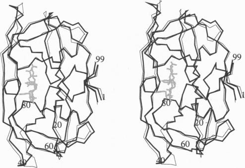
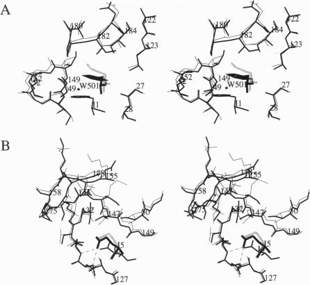
The crystal structure of HIV-2 protease in complex with a reduced amide inhibitor [BI-LA-398; Phe-Val-Phe-psi (CH2NH)-Leu-Glu-Ile-amide] has been determined at 2.2-A resolution and refined to a crystallographic R factor of 17.6%. The rms deviation from ideality in bond lengths is 0.018 A and in bond angles is 2.8 degrees. The largest structural differences between HIV-1 and HIV-2 proteases are located at residues 15-20, 34-40, and 65-73, away from the flap region and the substrate binding sites. The rms distance between equivalent C alpha atoms of HIV-1 and HIV-2 protease structures excluding these residues is 0.5 A. The shapes of the S1 and S2 pockets in the presence of this inhibitor are essentially unperturbed by the amino acid differences between HIV-1 and HIV-2 proteases. The interaction of the inhibitor with HIV-2 protease is similar to that observed in HIV-1 protease structures. The unprotected N terminus of the inhibitor interacts with the side chains of Asp-29 and Asp-30. The glutamate side chain of the inhibitor forms hydrogen bonds with the main-chain amido groups of residues 129 and 130.
已确定与一种还原酰胺抑制剂[BI-LA-398;苯丙氨酸-缬氨酸-苯丙氨酸-ψ(CH2NH)-亮氨酸-谷氨酸-异亮氨酸-酰胺]结合的HIV-2蛋白酶的晶体结构,分辨率为2.2埃,并精修至晶体学R因子为17.6%。键长与理想值的均方根偏差为0.018埃,键角为2.8度。HIV-1和HIV-2蛋白酶之间最大的结构差异位于15-20、34-40和65-73位残基处,远离侧翼区和底物结合位点。排除这些残基后,HIV-1和HIV-2蛋白酶结构中等效Cα原子之间的均方根距离为0.5埃。在存在这种抑制剂的情况下,S1和S2口袋的形状基本上不受HIV-1和HIV-2蛋白酶之间氨基酸差异的干扰。抑制剂与HIV-2蛋白酶的相互作用与在HIV-1蛋白酶结构中观察到的相似。抑制剂未受保护的N末端与天冬氨酸-29和天冬氨酸-30的侧链相互作用。抑制剂的谷氨酸侧链与129和130位残基的主链酰胺基团形成氢键。