Brown L F, Lanir N, McDonagh J, Tognazzi K, Dvorak A M, Dvorak H F
Department of Pathology, Beth Israel Hospital, Boston, MA 02215.
Am J Pathol. 1993 Jan;142(1):273-83.
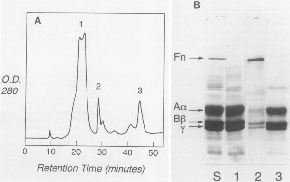
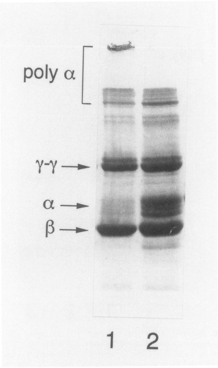
In healing wounds and many solid tumors, locally increased microvascular permeability results in extravasation of fibrinogen and its extravascular coagulation to form a fibrin gel, with concomitant covalent cross-linking of fibrin by factor XIIIa. Subsequently, inflammatory cells, fibroblasts, and endothelial cells migrate into the gel and organize it into granulation tissue and later into mature collagenous connective tissue. To gain insight into some of the cell migration events associated with these processes, we developed a quantitative in vitro assay that permits the study of fibroblast migration in fibrin gels. Early passage human or rat fibroblasts were allowed to attach to tissue culture dishes and then were overlaid with a thin layer of fibrinogen that was clotted with thrombin. Fibroblasts began to migrate upwards into the fibrin within 24 hours and their numbers and the distance migrated were quantified over several days. The extent of fibroblast migration was affected importantly by the nature of the fibrin clot. Fibroblasts migrated optimally into gels prepared from fibrinogen at concentrations of -3 mg/ml; ie, near normal plasma fibrinogen levels. Migration was greatly enhanced by extensive cross-linking of the fibrin alpha-chains by factor XIIIa, as occurs when clotting takes place in vivo. When fibrinogen was clotted in Dulbecco's modified Eagle's medium, gamma-chains were cross-linked, but alpha-chain cross-linking was strikingly inhibited, and fibroblasts migrated poorly. Gels prepared from factor XIII-depleted fibrinogen exhibited neither alpha-nor gamma-chain cross-linking and did not support fibroblast migration. Further purification of fibrinogen by anion exchange high pressure liquid chromatography depleted fibrinogen of fibronectin, plasminogen, and other impurities; this purified fibrinogen clotted to form fibrin gels that supported reproducible fibroblast migration.
在愈合伤口和许多实体肿瘤中,局部微血管通透性增加会导致纤维蛋白原外渗及其在血管外凝固形成纤维蛋白凝胶,同时因子XIIIa会使纤维蛋白发生共价交联。随后,炎症细胞、成纤维细胞和内皮细胞迁移到凝胶中,并将其组织成肉芽组织,随后再形成成熟的胶原结缔组织。为了深入了解与这些过程相关的一些细胞迁移事件,我们开发了一种定量体外试验,该试验可用于研究成纤维细胞在纤维蛋白凝胶中的迁移。将早期传代的人或大鼠成纤维细胞接种到组织培养皿上,然后覆盖一层用凝血酶凝结的纤维蛋白原薄层。成纤维细胞在24小时内开始向上迁移到纤维蛋白中,并在数天内对其数量和迁移距离进行定量。成纤维细胞的迁移程度受到纤维蛋白凝块性质的重要影响。成纤维细胞最佳地迁移到由浓度为-3mg/ml的纤维蛋白原制备的凝胶中;即接近正常血浆纤维蛋白原水平。如在体内凝血时发生的那样,因子XIIIa对纤维蛋白α链的广泛交联极大地增强了迁移。当纤维蛋白原在杜尔贝科改良伊格尔培养基中凝结时,γ链会发生交联,但α链交联受到显著抑制,成纤维细胞迁移较差。由缺乏因子XIII的纤维蛋白原制备的凝胶既不显示α链也不显示γ链交联,并且不支持成纤维细胞迁移。通过阴离子交换高压液相色谱对纤维蛋白原进行进一步纯化,去除了纤维连接蛋白、纤溶酶原和其他杂质;这种纯化的纤维蛋白原凝结形成支持可重复的成纤维细胞迁移的纤维蛋白凝胶。