Kaul D K, Fabry M E, Costantini F, Rubin E M, Nagel R L
Division of Hematology, Albert Einstein College of Medicine, New York 10461, USA.
J Clin Invest. 1995 Dec;96(6):2845-53. doi: 10.1172/JCI118355.
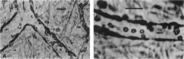
Intravascular sickling, red cell-endothelium interaction, and altered microvascular responses have been suggested to contribute to the pathophysiology of human sickle cell disease, but have never been demonstrated under in vivo flow. To address this issue, we have examined a transgenic mouse line, alphaHbetaSbetaS-Antilles [betaMDD] which has a combined high (78%) expression of beta S and beta S-Antilles globins. In vivo microcirculatory studies using the cremaster muscle preparation showed adhesion of red cells, restricted to postcapillary venules, in transgenic mice but not in control mice. Electron microscopy revealed distinct contacts between the red cell membrane and the endothelium surface. Some red cells exhibiting sickling were regularly observed in the venular flow. Infusion of transgenic mouse red cells into the ex vivo mesocecum vasculature also showed adhesion of mouse red cells exclusively in venules. Under resting conditions (pO2, 15-20 mmHg), there were no differences in the cremaster microvascular diameters of control and transgenic mice; however, transgenic mice showed a drastic reduction in microvascular red cell velocities (Vrbc) with maximal Vrbc decrease (> 60%) occurring in venules, the sites of red cell adhesion and sickling. Local, transient hyperoxia (pO2, 150 mmHg) resulted in striking differences between control and transgenic mice. In controls, oxygen caused a 69% arteriolar constriction, accompanied by 75% reduction in Vrbc. In contrast, in transgenic mice, hyperoxia resulted in only 8% decrease in the arteriolar diameter and in 68% increase in VrBC; the latter is probably due to an improved flow behavior of red cells as a consequence of unsickling. In summary, the high expression of human sickle hemoglobin in the mouse results not only in intravascular sickling but also red cell-endothelium interaction. The altered microvascular response to oxygen could be secondary to blood rheological changes, although possible intrinsic differences in the endothelial cell/vascular smooth muscle function in the transgenic mouse may also contribute. These sickle transgenic mice could serve as a useful model to investigate vasoocclusive mechanisms, as well as to test potential therapies.
血管内镰变、红细胞与内皮细胞的相互作用以及微血管反应改变被认为与人类镰状细胞病的病理生理学有关,但从未在体内血流情况下得到证实。为了解决这个问题,我们研究了一种转基因小鼠品系,αHβSβS-安的列斯[βMDD],其βS和βS-安的列斯球蛋白的表达量较高(78%)。使用提睾肌制备进行的体内微循环研究显示,转基因小鼠的红细胞黏附仅限于毛细血管后微静脉,而对照小鼠则未出现这种情况。电子显微镜显示红细胞膜与内皮细胞表面有明显接触。在微静脉血流中经常观察到一些呈现镰变的红细胞。将转基因小鼠的红细胞注入离体的盲肠系膜血管系统中,也显示小鼠红细胞仅在微静脉中黏附。在静息状态下(pO2为15 - 20 mmHg),对照小鼠和转基因小鼠的提睾肌微血管直径没有差异;然而,转基因小鼠的微血管红细胞速度(Vrbc)急剧降低,最大Vrbc降低(>60%)发生在红细胞黏附和镰变的微静脉部位。局部短暂性高氧(pO2为150 mmHg)导致对照小鼠和转基因小鼠之间出现显著差异。在对照小鼠中,氧气导致小动脉收缩69%,同时Vrbc降低75%。相比之下,在转基因小鼠中,高氧导致小动脉直径仅降低8%,而VrBC增加68%;后者可能是由于红细胞去镰变后血流行为改善所致。总之,小鼠中人类镰状血红蛋白的高表达不仅导致血管内镰变,还导致红细胞与内皮细胞相互作用。对氧气的微血管反应改变可能继发于血液流变学变化,尽管转基因小鼠内皮细胞/血管平滑肌功能可能存在的内在差异也可能起作用。这些镰状转基因小鼠可作为研究血管闭塞机制以及测试潜在治疗方法的有用模型。