Sunness J S, Applegate C A, Haselwood D, Rubin G S
Lions Vision Center, Wilmer Ophthalmological Institute, Johns Hopkins University School of Medicine, Baltimore, USA.
Ophthalmology. 1996 Sep;103(9):1458-66. doi: 10.1016/s0161-6420(96)30483-1.
To study fixation patterns and reading rates in eyes with central scotomas from geographic atrophy (GA) of age-related macular degeneration and to compare fixation patterns with those of patients with Stargardt disease.
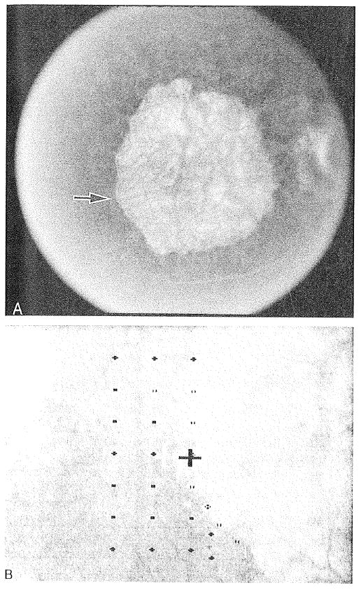
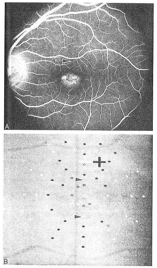
Scanning laser ophthalmoscope analysis of fixation patterns in eyes with 20/80 to 20/200 visual acuity. Included were 41 eyes of 35 patients with GA and 10 eyes of 5 patients with Stargardt disease. The patients with GA also were tested for maximum reading rate, and the size of the areas of atrophy were measured by fundus photograph analysis.
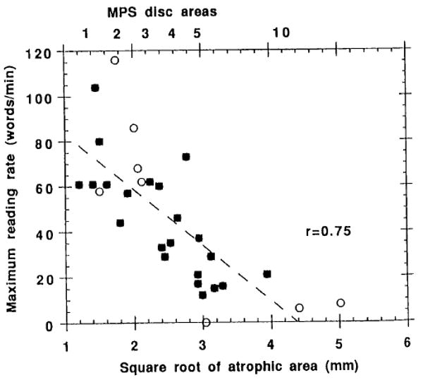
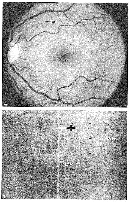
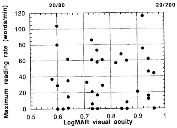
Sixty-three percent of GA eyes fixating outside the atrophy placed the scotoma to the right of fixation in visual field space, 22% placed the scotoma above fixation, and 15% placed it to the left, regardless of the laterality of the GA eye. Fixation was stable in subsequent years of testing for scotoma placement to the right of or above fixation. All GA eyes fixated immediately adjacent to the atrophy. In contrast, seven of ten eyes with Stargardt disease fixated at a considerable distance from the scotoma border, with the dense scotoma far above the fixation site in visual field space. For the patients with GA, the maximum reading rate was highly correlated with size of the atrophic area, but not with age or visual acuity within the limited visual acuity range tested. There was a trend to more rapid reading with the scotoma above fixation and slower reading with the scotoma to the left.
There is a preference for fixation with the scotoma to the right in eyes with GA. Patients with Stargardt disease use different strategies for fixation, perhaps due to subclinical pathology adjacent to the atrophic regions. The size of the atrophic area in GA plays the predominant role in reading rate for eyes that have already lost foveal vision.
研究年龄相关性黄斑变性地理性萎缩(GA)所致中心暗点患者的注视模式和阅读速度,并将其注视模式与Stargardt病患者的进行比较。
对视力为20/80至20/200的眼睛进行扫描激光检眼镜注视模式分析。纳入35例GA患者的41只眼睛和5例Stargardt病患者的10只眼睛。对GA患者还进行了最大阅读速度测试,并通过眼底照片分析测量萎缩区域的大小。
63%的GA眼睛在萎缩区域外注视时,暗点在视野空间中位于注视点右侧,22%的暗点位于注视点上方,15%位于左侧,与GA眼睛的左右侧无关。在随后几年对位于注视点右侧或上方的暗点位置进行测试时,注视是稳定的。所有GA眼睛都紧邻萎缩区域注视。相比之下,10只Stargardt病眼睛中有7只在距暗点边界相当远的位置注视,致密暗点在视野空间中远高于注视点。对于GA患者,最大阅读速度与萎缩区域大小高度相关,但在所测试的有限视力范围内与年龄或视力无关。暗点在注视点上方时阅读速度有加快趋势,暗点在左侧时阅读速度较慢。
GA患者的眼睛倾向于将暗点置于右侧进行注视。Stargardt病患者采用不同的注视策略,可能是由于萎缩区域附近的亚临床病理改变。GA中萎缩区域的大小在已经丧失中央凹视力的眼睛的阅读速度中起主要作用。