Parasassi T, Gratton E, Yu W M, Wilson P, Levi M
Istituto di Medicina Sperimentale, CNR, Rome, Italy.
Biophys J. 1997 Jun;72(6):2413-29. doi: 10.1016/S0006-3495(97)78887-8.
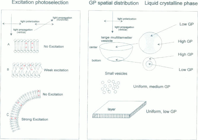
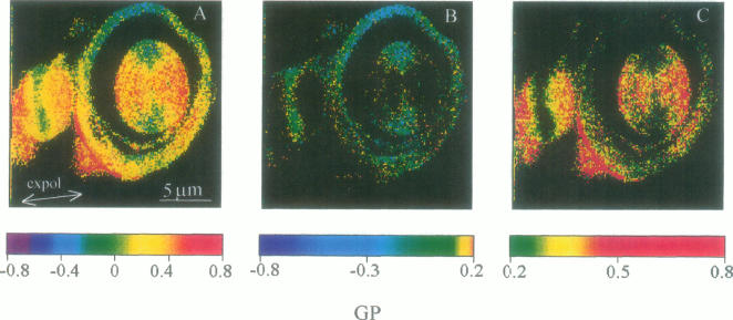
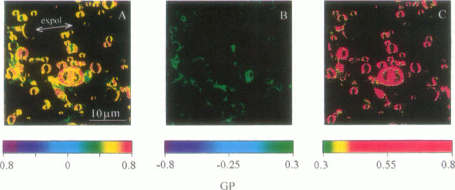
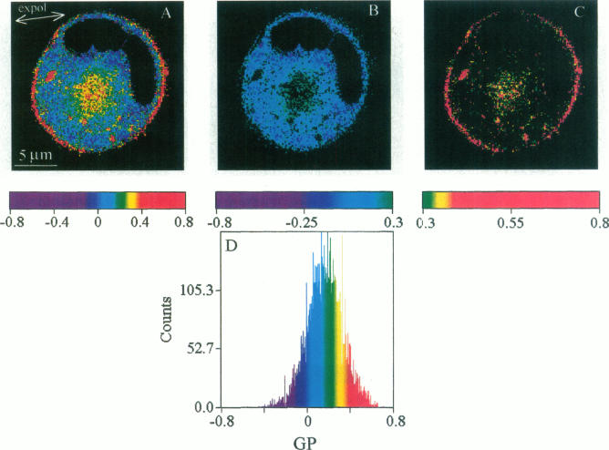
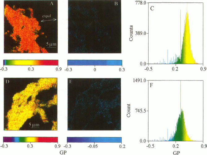
Two-photon excitation microscopy shows coexisting regions of different generalized polarization (GP) in phospholipid vesicles, in red blood cells, in a renal tubular cell line, and in purified renal brushborder and basolateral membranes labeled with the fluorescent probe laurdan. The GP function measures the relative water content of the membrane. In the present study we discuss images obtained with polarized laser excitation, which selects different molecular orientations of the lipid bilayer corresponding to different spatial regions. The GP distribution in the gel-phase vesicles is relatively narrow, whereas the GP distribution in the liquid-crystalline phase vesicles (DOPC and DLPC) is broad. Analysis of images obtained with polarized excitation of the liquid-crystalline phase vesicles leads to the conclusion that coexisting regions of different GP must have dimensions smaller than the microscope resolution (approximately 200 nm radially and 600 nm axially). Vesicles of an equimolar mixture of DOPC and DPPC show coexisting rigid and fluid domains (high GP and low GP), but the rigid domains, which are preferentially excited by polarized light, have GP values lower than the pure gel-phase domains. Cholesterol strongly modifies the domain morphology. In the presence of 30 mol% cholesterol, the broad GP distribution of the DOPC/DPPC equimolar sample becomes narrower. The sample is still very heterogeneous, as demonstrated by the separations of GP disjoined regions, which are the result of photoselection of regions of different lipid orientation. In intact red blood cells, microscopic regions of different GP can be resolved, whereas in the renal cells GP domains have dimensions smaller than the microscope resolution. Preparations of renal apical brush border membranes and basolateral membranes show well-resolved GP domains, which may result from a different local orientation, or the domains may reflect a real heterogeneity of these membranes.
双光子激发显微镜显示,在磷脂囊泡、红细胞、肾小管细胞系以及用荧光探针劳丹标记的纯化肾刷状缘和基底外侧膜中,存在不同广义极化(GP)的共存区域。GP功能测量膜的相对含水量。在本研究中,我们讨论了用偏振激光激发获得的图像,该激发选择了与不同空间区域相对应的脂质双层的不同分子取向。凝胶相囊泡中的GP分布相对较窄,而液晶相囊泡(DOPC和DLPC)中的GP分布较宽。对液晶相囊泡偏振激发获得的图像分析得出结论,不同GP的共存区域的尺寸必须小于显微镜分辨率(径向约200nm,轴向约600nm)。DOPC和DPPC等摩尔混合物的囊泡显示出刚性和流体域共存(高GP和低GP),但优先被偏振光激发的刚性域的GP值低于纯凝胶相域。胆固醇强烈改变域形态。在存在30mol%胆固醇的情况下,DOPC/DPPC等摩尔样品的宽GP分布变窄。如GP分离区域所示,样品仍然非常不均匀,这是不同脂质取向区域光选择的结果。在完整的红细胞中,可以分辨出不同GP的微观区域,而在肾细胞中,GP域的尺寸小于显微镜分辨率。肾顶端刷状缘膜和基底外侧膜的制剂显示出分辨良好的GP域,这可能是由于不同的局部取向,或者这些域可能反映了这些膜的真正异质性。