Dubrey S W, Cha K, Skinner M, LaValley M, Falk R H
Section of Cardiology, Boston University School of Medicine, Massachusetts, USA.
Heart. 1997 Jul;78(1):74-82. doi: 10.1136/hrt.78.1.74.
To determine whether patients with myocardial amyloidosis due either to AL (primary) amyloid or familial amyloid have distinguishing echocardiographic or electrocardiographic features; and to compare the prevalence of heart failure and survival in the two types of amyloidosis in relation to echocardiographic findings.
Blinded group comparison of randomly selected cases of cardiac amyloidosis.
International referral centre for amyloid research and treatment.
36 patients with cardiac amyloid heart disease, of whom 12 had familial and 24 had primary AL amyloidosis.
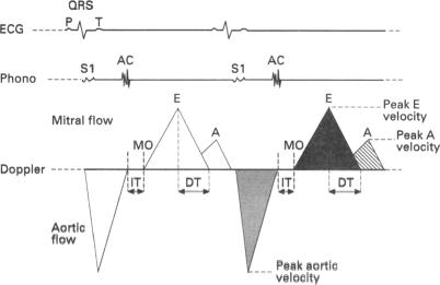
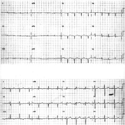
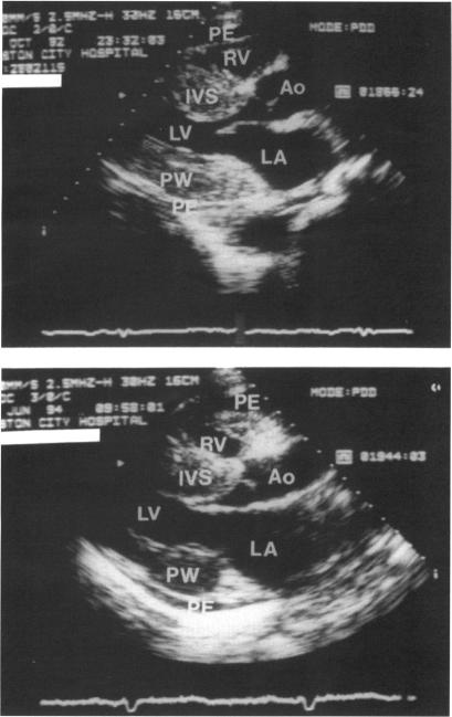
Familial and AL echocardiograms were morphologically indistinguishable, with similar left ventricular wall thickness, mean (SD) 15.4 (2.3) nu 15.8 (2.5) mm, respectively; right ventricular wall thickness was also similar between amyloid types: 9.6 (2.8) nu 9.7 (6.5) mm, respectively. Doppler indices of left and right ventricular function, left ventricular volume, and ejection fraction were also similar. Low voltage electrocardiograms (< 0.5 mV) were more common in the AL (16/24, 67%) than in the familial group (4/12, 25%), P < 0.05. The one year survival for familial and AL forms was 92% (11/12) nu 38% (6/24), respectively, with virtually all deaths due to cardiac causes.
Although cardiac involvement is echocardiographically indistinguishable, cardiac mortality is very different between the two forms of amyloidosis. Preservation of electrocardiographic voltage in familial amyloidosis suggests that the particular biochemical characteristics of distinct types of amyloid fibril have different pathological effects on the myocardium. This distinction becomes critical in the evaluation, treatment, and management of patients who have a diagnosis within the spectrum of the protein deposition diseases.
确定由AL(原发性)淀粉样蛋白或家族性淀粉样蛋白引起的心肌淀粉样变性患者是否具有独特的超声心动图或心电图特征;并根据超声心动图结果比较两种类型淀粉样变性中心力衰竭的患病率和生存率。
对随机选择的心脏淀粉样变性病例进行盲法分组比较。
国际淀粉样变性研究与治疗转诊中心。
36例心脏淀粉样变性心脏病患者,其中12例为家族性,24例为原发性AL淀粉样变性。
家族性和AL型超声心动图在形态上无法区分,左心室壁厚度相似,分别为平均(标准差)15.4(2.3)毫米和15.8(2.5)毫米;淀粉样变性类型之间的右心室壁厚度也相似:分别为9.6(2.8)毫米和9.7(6.5)毫米。左、右心室功能、左心室容积和射血分数的多普勒指标也相似。低电压心电图(<0.5mV)在AL组(16/24,67%)比家族性组(4/12,25%)更常见,P<0.05。家族性和AL型的一年生存率分别为92%(11/12)和38%(6/24),几乎所有死亡均由心脏原因导致。
尽管心脏受累在超声心动图上无法区分,但两种形式的淀粉样变性中心脏死亡率差异很大。家族性淀粉样变性中保留心电图电压表明,不同类型淀粉样纤维的特定生化特征对心肌有不同的病理影响。这种区别在对蛋白质沉积疾病范围内诊断的患者进行评估、治疗和管理时变得至关重要。