Conti L, Rainaldi G, Matarrese P, Varano B, Rivabene R, Columba S, Sato A, Belardelli F, Malorni W, Gessani S
Laboratory of Virology, Istituto Superiore di Sanità, Viale Regina Elena, 299-00161 Rome, Italy.
J Exp Med. 1998 Feb 2;187(3):403-13. doi: 10.1084/jem.187.3.403.
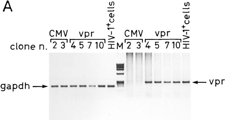
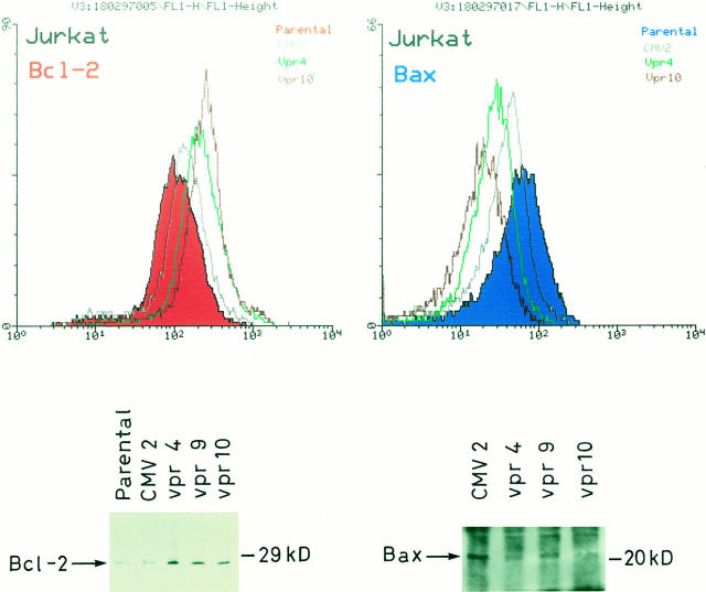
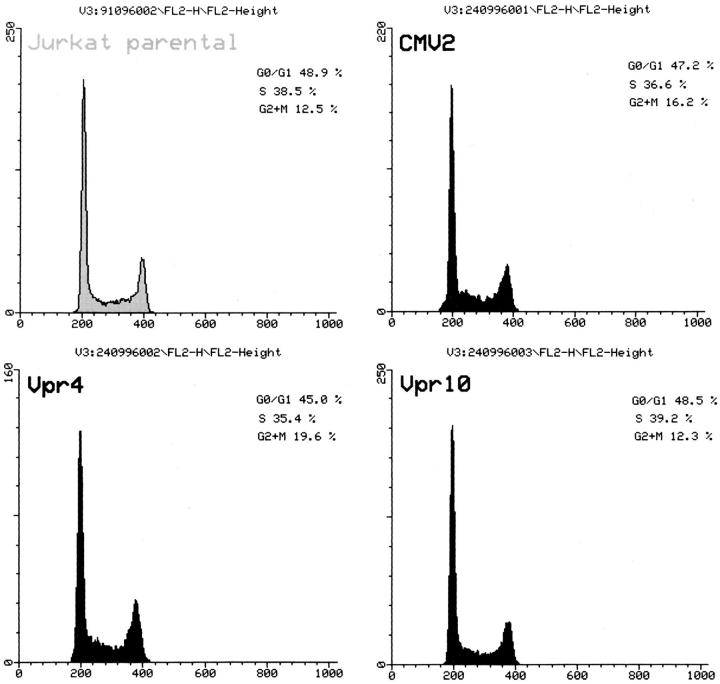
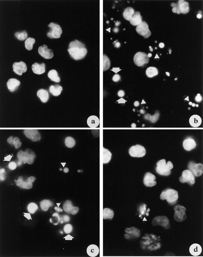
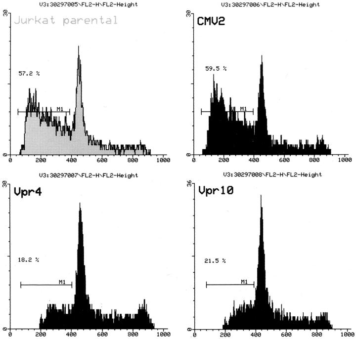
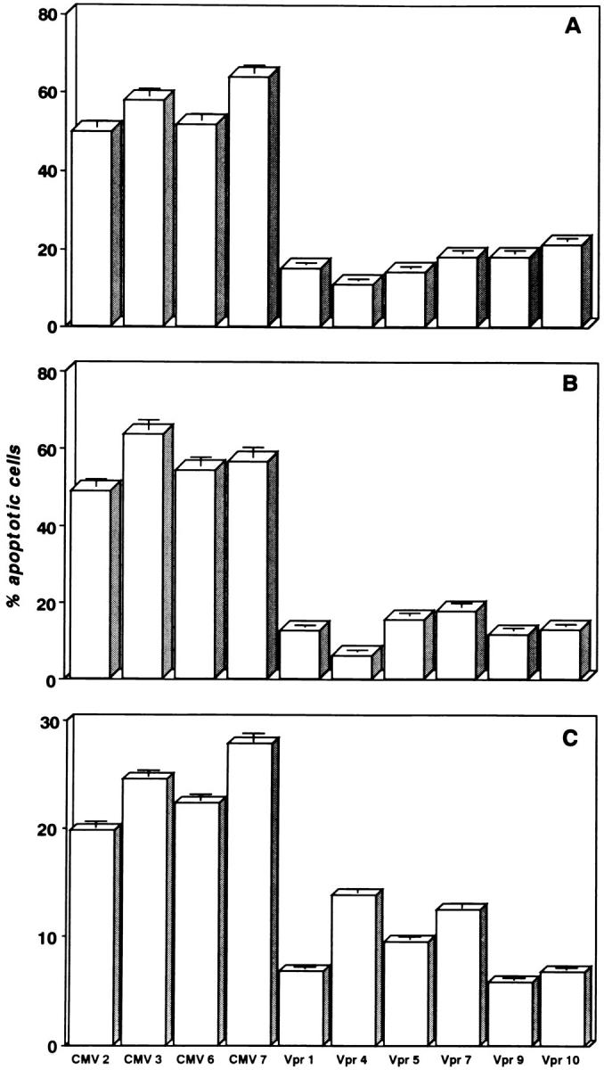
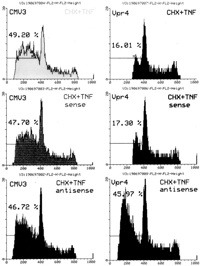
Although apoptosis is considered one of the major mechanisms of CD4(+) T cell depletion in HIV-infected patients, the virus-infected cells somehow appear to be protected from apoptosis, which generally occurs in bystander cells. Vpr is an auxiliary HIV-1 protein, which, unlike the other regulatory gene products, is present at high copy number in virus particles. We established stable transfectants of CD4+ T Jurkat cells constitutively expressing low levels of vpr. These clones exhibited cell cycle characteristics similar to those of control-transfected cells. Treatment of control clones with apoptotic stimuli (i.e., cycloheximide/tumor necrosis factor alpha (TNF-alpha), anti-Fas antibody, or serum starvation) resulted in a massive cell death by apoptosis. In contrast, all the vpr-expressing clones showed an impressive protection from apoptosis independently of the inducer. Notably, vpr antisense phosphorothioate oligodeoxynucleotides render vpr-expressing cells as susceptible to apoptosis induced by cycloheximide and TNF-alpha as the control clones. Moreover, the constitutive expression of HIV-1 vpr resulted in the upregulation of bcl-2, an oncogene endowed with antiapoptotic activities, and in the downmodulation of bax, a proapoptotic factor of the bcl-2 family. Altogether, these results suggest that low levels of the endogenous vpr protein can interfere with the physiological turnover of T lymphocytes at early stages of virus infection, thus facilitating HIV persistence and, subsequently, viral spread. This might explain why apoptosis mostly occurs in bystander uninfected cells in AIDS patients.
尽管细胞凋亡被认为是HIV感染患者CD4(+) T细胞耗竭的主要机制之一,但病毒感染的细胞似乎以某种方式受到保护而免于凋亡,而凋亡通常发生在旁观者细胞中。Vpr是一种辅助性HIV-1蛋白,与其他调节基因产物不同,它以高拷贝数存在于病毒颗粒中。我们构建了稳定转染的组成型表达低水平vpr的CD4+ T Jurkat细胞。这些克隆表现出与对照转染细胞相似的细胞周期特征。用凋亡刺激物(即放线菌酮/肿瘤坏死因子α(TNF-α)、抗Fas抗体或血清饥饿)处理对照克隆会导致大量细胞通过凋亡而死亡。相比之下,所有表达vpr的克隆均表现出对凋亡的显著保护作用,且与诱导剂无关。值得注意的是,vpr反义硫代磷酸酯寡脱氧核苷酸使表达vpr的细胞对放线菌酮和TNF-α诱导的凋亡与对照克隆一样敏感。此外,HIV-1 vpr的组成型表达导致具有抗凋亡活性的癌基因bcl-2上调,以及bcl-2家族的促凋亡因子bax下调。总之,这些结果表明,内源性vpr蛋白的低水平可在病毒感染早期干扰T淋巴细胞的生理性更新,从而促进HIV的持续存在以及随后的病毒传播。这可能解释了为什么细胞凋亡大多发生在艾滋病患者的未感染旁观者细胞中。