Shoji M, Hancock W W, Abe K, Micko C, Casper K A, Baine R M, Wilcox J N, Danave I, Dillehay D L, Matthews E, Contrino J, Morrissey J H, Gordon S, Edgington T S, Kudryk B, Kreutzer D L, Rickles F R
Hematologic Disease Branch, DASTLR/NCID, Centers for Disease Control and Prevention, Atlanta, Georgia 30333, USA.
Am J Pathol. 1998 Feb;152(2):399-411.
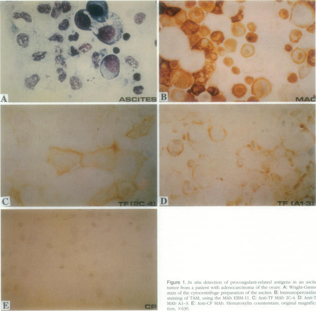
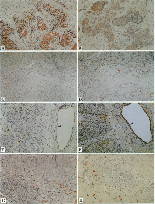
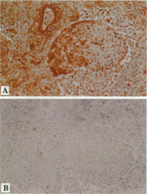
Thrombin-catalyzed, cross-linked fibrin (XLF) formation is a characteristic histopathological finding in many human and experimental tumors and is thought to be of importance in the local host defense response. Although the pathogenesis of tumor-associated fibrin deposition is not entirely clear, several tumor procoagulants have been described as likely primary stimuli for the generation of thrombin (and XLF) in the tumor microenvironment (TME). In a previous study of a variety of human tumors we have shown that tissue factor (TF) is the major procoagulant. However, the relative contribution to fibrin deposition in the TME of tumor cell TF and host cell TF (eg, macrophage-derived) was not established. In addition, recent evidence has implicated TF in the regulation of the synthesis of the pro-angiogenic factor vascular endothelial growth factor (VEGF) by tumor cells. In the current study we used in situ techniques to determine the cellular localization of XLF, TF, VEGF, and an alternative tumor procoagulant, so-called cancer procoagulant (CP), a cysteine protease that activates clotting factor X. In lung cancer we have found XLF localized predominantly to the surface of tumor-associated macrophages, as well as to some endothelial cells and perivascular fibroblasts in the stromal area of the tumors co-distributed with TF at the interface of the tumor and host cells. Cancer pro-coagulant was localized to tumor cells in several cases but not in conjunction with the deposition of XLF. TF and VEGF were co-localized in both lung cancer and breast cancer cells by in situ hybridization and immunohistochemical staining. Furthermore, a strong relationship was found between the synthesis of TF and VEGF levels in human breast cancer cell lines (r2 = 0.84; P < 0.0001). Taken together, these data are consistent with a highly complex interaction between tumor cells, macrophages, and endothelial cells in the TME leading to fibrin formation and tumor angiogenesis.
凝血酶催化的交联纤维蛋白(XLF)形成是许多人类和实验性肿瘤的特征性组织病理学表现,被认为在局部宿主防御反应中具有重要意义。尽管肿瘤相关纤维蛋白沉积的发病机制尚不完全清楚,但几种肿瘤促凝剂已被描述为肿瘤微环境(TME)中凝血酶(和XLF)产生的可能主要刺激因素。在先前对多种人类肿瘤的研究中,我们已经表明组织因子(TF)是主要的促凝剂。然而,肿瘤细胞TF和宿主细胞TF(例如巨噬细胞衍生的)对TME中纤维蛋白沉积的相对贡献尚未确定。此外,最近的证据表明TF参与了肿瘤细胞对促血管生成因子血管内皮生长因子(VEGF)合成的调节。在当前的研究中,我们使用原位技术来确定XLF(交联纤维蛋白)、TF、VEGF以及另一种肿瘤促凝剂(所谓的癌促凝剂,CP,一种激活凝血因子X的半胱氨酸蛋白酶)的细胞定位。在肺癌中,我们发现XLF主要定位于肿瘤相关巨噬细胞的表面,以及肿瘤基质区域的一些内皮细胞和血管周围成纤维细胞,在肿瘤与宿主细胞的界面处与TF共同分布。在一些病例中,癌促凝剂定位于肿瘤细胞,但与XLF的沉积无关。通过原位杂交和免疫组织化学染色,TF和VEGF在肺癌和乳腺癌细胞中共同定位。此外,在人乳腺癌细胞系中发现TF的合成与VEGF水平之间存在很强的相关性(r2 = 0.84;P < 0.0001)。综上所述,这些数据与TME中肿瘤细胞、巨噬细胞和内皮细胞之间导致纤维蛋白形成和肿瘤血管生成的高度复杂相互作用一致。