McCracken J S, Klintworth G K
Am J Pathol. 1976 Oct;85(1):167-82.
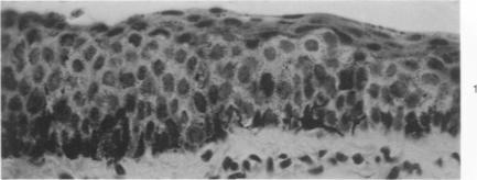
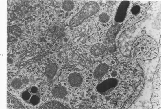
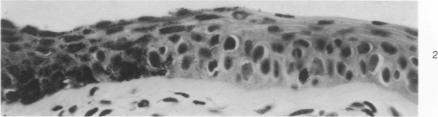
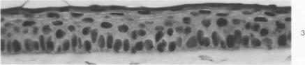
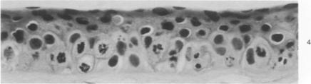
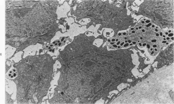
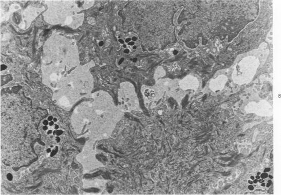
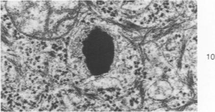
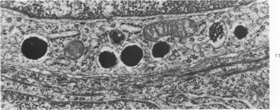
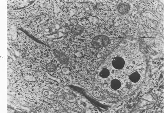
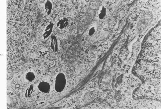
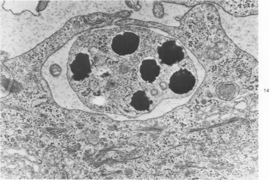
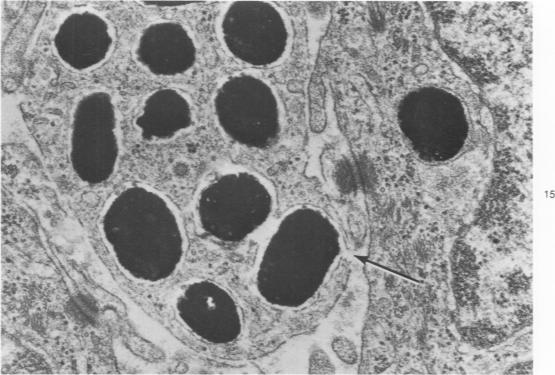
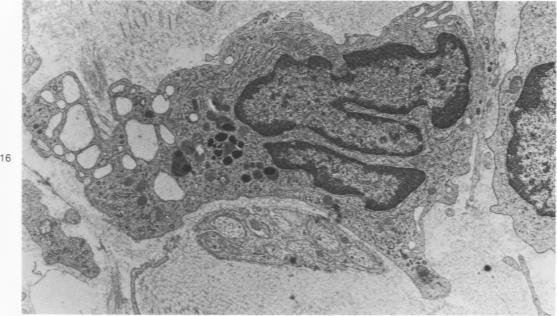
Melanin pigmentation of the corneal epithelium was induced in pigmented guinea pigs by the topical application of colchicine to their eyes or by corneal cauterization with silver nitrate. With colchicine the pigmentation was preceded by the development of an abnormal corneal epithelium in which numerous cells became arrested in cell division. The corneal melanosis resulted largely from the migration of melanocytes into the corneal epithelium from the normally pigmented contiguous conjunctiva and to a lesser extent from the presence of melanin granules within corneal epithelial cells. In both models a leukocytic and vascular invasion of the cornea proceded and accompanied the migration of melanocytes into the corneal epithelium. Electron microscopy disclosed cells with the same morphology as conjunctival melanocytes between the epithelial cells of the cornea. Mature melanin granules were also present within some squamous epithelial cells as individual granules or as clusters. The ultrastructural findings are viewed in relation to how melanin granules are transferred from melanocytes to epithelial cells. Evidence is presented which suggests that malanin granule transfer may follow the fusion of the membranes of the melanocytes and epithelial cells.
通过向有色豚鼠的眼睛局部应用秋水仙碱或用硝酸银进行角膜烧灼,可诱导角膜上皮的黑色素沉着。使用秋水仙碱时,色素沉着之前会出现异常的角膜上皮,其中许多细胞在细胞分裂中停滞。角膜黑变病主要是由于黑素细胞从正常色素沉着的相邻结膜迁移到角膜上皮,在较小程度上是由于角膜上皮细胞内存在黑色素颗粒。在这两种模型中,角膜均出现白细胞和血管浸润,并伴随黑素细胞迁移到角膜上皮。电子显微镜检查发现角膜上皮细胞之间存在与结膜黑素细胞形态相同的细胞。成熟的黑色素颗粒也以单个颗粒或簇的形式存在于一些鳞状上皮细胞内。结合黑色素颗粒如何从黑素细胞转移到上皮细胞来观察超微结构的发现。有证据表明,黑色素颗粒转移可能发生在黑素细胞和上皮细胞膜融合之后。