McGowan K M, Coulombe P A
Department of Biological Chemistry and Department of Dermatology, The Johns Hopkins University School of Medicine, Baltimore, Maryland 21205, USA.
J Cell Biol. 1998 Oct 19;143(2):469-86. doi: 10.1083/jcb.143.2.469.
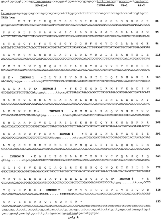
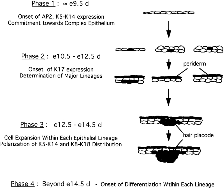
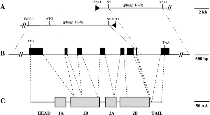
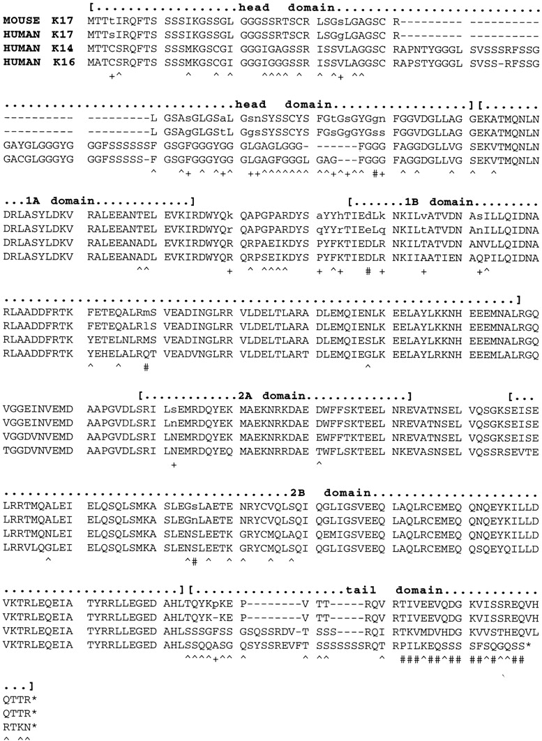
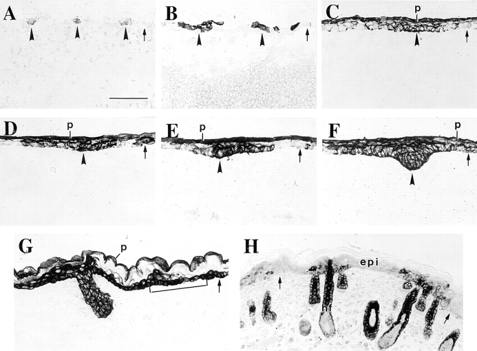
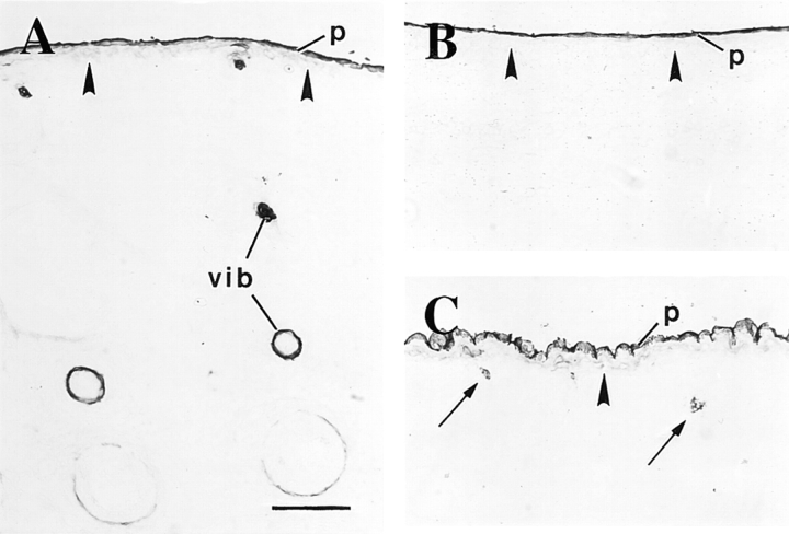
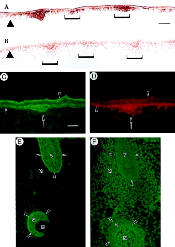
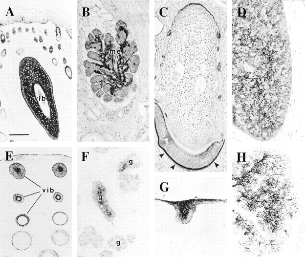
The type I keratin 17 (K17) shows a peculiar localization in human epithelial appendages including hair follicles, which undergo a growth cycle throughout adult life. Additionally K17 is induced, along with K6 and K16, early after acute injury to human skin. To gain further insights into its potential function(s), we cloned the mouse K17 gene and investigated its expression during skin development. Synthesis of K17 protein first occurs in a subset of epithelial cells within the single-layered, undifferentiated ectoderm of embryonic day 10.5 mouse fetuses. In the ensuing 48 h, K17-expressing cells give rise to placodes, the precursors of ectoderm-derived appendages (hair, glands, and tooth), and to periderm. During early development, there is a spatial correspondence in the distribution of K17 and that of lymphoid-enhancer factor (lef-1), a DNA-bending protein involved in inductive epithelial-mesenchymal interactions. We demonstrate that ectopic lef-1 expression induces K17 protein in the skin of adult transgenic mice. The pattern of K17 gene expression during development has direct implications for the morphogenesis of skin epithelia, and points to the existence of a molecular relationship between development and wound repair.
I型角蛋白17(K17)在包括毛囊在内的人类上皮附属器中呈现出特殊的定位,毛囊在成年期会经历一个生长周期。此外,在人类皮肤急性损伤后早期,K17与K6和K16一起被诱导表达。为了进一步深入了解其潜在功能,我们克隆了小鼠K17基因,并研究了其在皮肤发育过程中的表达情况。K17蛋白的合成首先出现在胚胎第10.5天小鼠胎儿单层未分化外胚层中的一部分上皮细胞中。在随后的48小时内,表达K17的细胞形成基板,即外胚层衍生附属器(毛发、腺体和牙齿)的前体,以及形成周皮。在早期发育过程中,K17的分布与淋巴增强因子(lef-1)的分布存在空间对应关系,lef-1是一种参与诱导性上皮-间充质相互作用的DNA弯曲蛋白。我们证明,异位表达lef-1可在成年转基因小鼠皮肤中诱导K17蛋白表达。K17基因在发育过程中的表达模式对皮肤上皮的形态发生具有直接影响,并表明发育与伤口修复之间存在分子关系。