• 文献检索
  • 文档翻译
  • 深度研究
  • 学术资讯
  • Suppr Zotero 插件Zotero 插件
  • 邀请有礼
  • 套餐&价格
  • 历史记录
应用&插件
Suppr Zotero 插件Zotero 插件浏览器插件Mac 客户端Windows 客户端微信小程序
定价
高级版会员购买积分包购买API积分包
服务
文献检索文档翻译深度研究API 文档MCP 服务
关于我们
关于 Suppr公司介绍联系我们用户协议隐私条款
关注我们

Suppr 超能文献

核心技术专利:CN118964589B侵权必究
粤ICP备2023148730 号-1Suppr @ 2026

文献检索

告别复杂PubMed语法,用中文像聊天一样搜索,搜遍4000万医学文献。AI智能推荐,让科研检索更轻松。

立即免费搜索

文件翻译

保留排版,准确专业,支持PDF/Word/PPT等文件格式,支持 12+语言互译。

免费翻译文档

深度研究

AI帮你快速写综述,25分钟生成高质量综述,智能提取关键信息,辅助科研写作。

立即免费体验

非霍奇金淋巴瘤的电子显微镜检查

Electron microscopy in the non-Hodgkin's lymphomata.

作者信息

Henry K

出版信息

Br J Cancer Suppl. 1975 Mar;2:73-93.

PMID:1101937
原文链接:https://pmc.ncbi.nlm.nih.gov/articles/PMC2149586/
Abstract

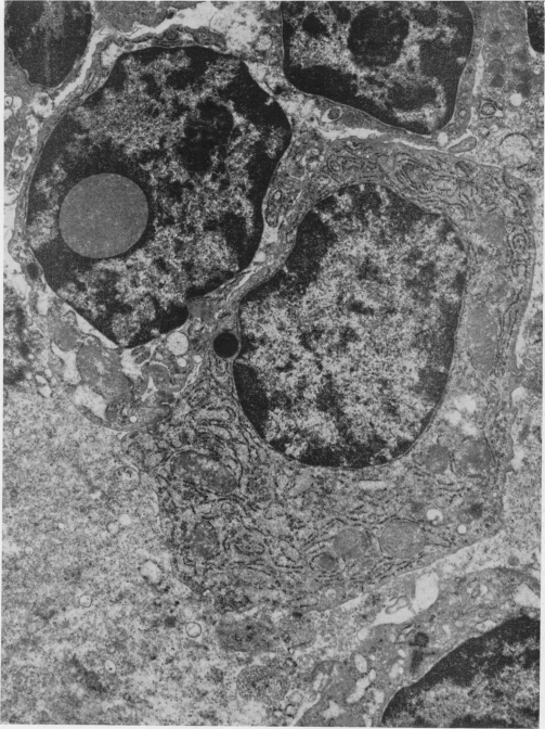
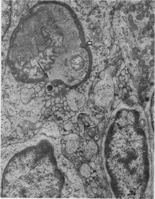
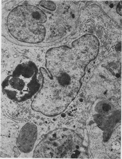
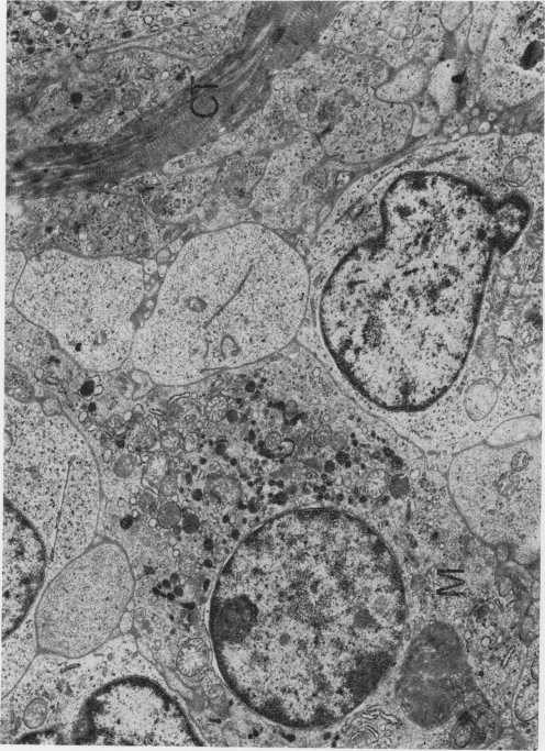
The component cells of peripheral lymphoid tissue have been divided into the lymphocyte and plasma cell lines, mononuclear phagocytic cells, dendritic "reticular cells", the reticular (supporting) cells and endothelial cells, and it is suggested that this system of cells should collectively be referred to as the lymphoreticular monoclear phagocyte system or LRMPS. Seventeen tumours of the LRMPS (excluding Hodgkin's disease) have been studied at ultrastructural level. Of these 17 non-Hodgkin lymphomata 5 were follicular lymphomata and 12 diffuse. It is concluded that electron microscopy plays a valuable role in the diagnosis of this group of tumours. Not only does it allow rejection of a diagnosis of lymphoma in certain anaplastic tumours, but it also enables a more precise identification of the cellular components of a lymphoma as well as indicating the degree of differentiation of the cell line involved. Additional advantages are the visualization of subcellular structures useful as markers, and by means of specialized immunoelectron microscopic techniques the identification of antigens and antibody formation within a given tumour. Two other results of this ultrastructural study are the indication that the dendritic cells of lymphoid follicles are derived from capillary endothelium, and the identification of certain anomalous formations derived from rough endoplasmic reticulum in the case of tumours showing plasmacytoid differentiation.

摘要

外周淋巴组织的组成细胞已被分为淋巴细胞系和浆细胞系、单核吞噬细胞、树突状“网状细胞”、网状(支持性)细胞和内皮细胞,有人建议这一细胞系统应统称为淋巴网状单核吞噬细胞系统或LRMPS。已在超微结构水平上研究了17例LRMPS肿瘤(不包括霍奇金病)。在这17例非霍奇金淋巴瘤中,5例为滤泡性淋巴瘤,12例为弥漫性淋巴瘤。得出的结论是,电子显微镜在这组肿瘤的诊断中发挥着重要作用。它不仅能在某些间变性肿瘤中排除淋巴瘤的诊断,还能更精确地识别淋巴瘤的细胞成分,并指出所涉及细胞系的分化程度。其他优点包括可观察到作为标志物的亚细胞结构,以及通过专门的免疫电子显微镜技术识别特定肿瘤内的抗原和抗体形成。这项超微结构研究的另外两个结果是,表明淋巴滤泡的树突状细胞起源于毛细血管内皮,以及在显示浆细胞样分化的肿瘤病例中识别出某些源自粗面内质网的异常结构。

https://cdn.ncbi.nlm.nih.gov/pmc/blobs/8b97/2149586/f2880fc1506a/brjcancersuppl00060-0098-a.jpg
https://cdn.ncbi.nlm.nih.gov/pmc/blobs/8b97/2149586/1cb510e9d3da/brjcancersuppl00060-0084-a.jpg
https://cdn.ncbi.nlm.nih.gov/pmc/blobs/8b97/2149586/43cd9c93d0bd/brjcancersuppl00060-0085-a.jpg
https://cdn.ncbi.nlm.nih.gov/pmc/blobs/8b97/2149586/734daa3c92f9/brjcancersuppl00060-0086-a.jpg
https://cdn.ncbi.nlm.nih.gov/pmc/blobs/8b97/2149586/18d31893779f/brjcancersuppl00060-0087-a.jpg
https://cdn.ncbi.nlm.nih.gov/pmc/blobs/8b97/2149586/2ba0e3f8d0f6/brjcancersuppl00060-0088-a.jpg
https://cdn.ncbi.nlm.nih.gov/pmc/blobs/8b97/2149586/a82b0201d8ed/brjcancersuppl00060-0089-a.jpg
https://cdn.ncbi.nlm.nih.gov/pmc/blobs/8b97/2149586/cf7b15789458/brjcancersuppl00060-0090-a.jpg
https://cdn.ncbi.nlm.nih.gov/pmc/blobs/8b97/2149586/f5252362b4e3/brjcancersuppl00060-0091-a.jpg
https://cdn.ncbi.nlm.nih.gov/pmc/blobs/8b97/2149586/c6c7572b282f/brjcancersuppl00060-0092-a.jpg
https://cdn.ncbi.nlm.nih.gov/pmc/blobs/8b97/2149586/84f108593b22/brjcancersuppl00060-0093-a.jpg
https://cdn.ncbi.nlm.nih.gov/pmc/blobs/8b97/2149586/c91d72ddbbe0/brjcancersuppl00060-0094-a.jpg
https://cdn.ncbi.nlm.nih.gov/pmc/blobs/8b97/2149586/a54e49040ca3/brjcancersuppl00060-0095-a.jpg
https://cdn.ncbi.nlm.nih.gov/pmc/blobs/8b97/2149586/b06b9771071e/brjcancersuppl00060-0096-a.jpg
https://cdn.ncbi.nlm.nih.gov/pmc/blobs/8b97/2149586/f8bb0ad1cdbf/brjcancersuppl00060-0097-a.jpg
https://cdn.ncbi.nlm.nih.gov/pmc/blobs/8b97/2149586/f2880fc1506a/brjcancersuppl00060-0098-a.jpg

相似文献

1
Electron microscopy in the non-Hodgkin's lymphomata.非霍奇金淋巴瘤的电子显微镜检查
Br J Cancer Suppl. 1975 Mar;2:73-93.
2
An ultrastructural analysis of lympho-reticular cell interactions in primary cultures of human non-lymphoid neoplasms and lymphomas.人非淋巴性肿瘤和淋巴瘤原代培养中淋巴网状细胞相互作用的超微结构分析
J Pathol. 1976 Oct;120(2):75-82. doi: 10.1002/path.1711200203.
3
Fascin, a sensitive new marker for Reed-Sternberg cells of hodgkin's disease. Evidence for a dendritic or B cell derivation?Fascin,一种霍奇金病里德-斯腾伯格细胞的新型敏感标志物。它来源于树突状细胞还是B细胞?
Am J Pathol. 1997 Feb;150(2):543-62.
4
Dendritic reticulum cell immunoreactivity for low-affinity nerve growth factor receptor in malignant lymphomas.恶性淋巴瘤中树突状网状细胞对低亲和力神经生长因子受体的免疫反应性。
Mod Pathol. 1996 Oct;9(10):959-65.
5
CD30 distribution. Immunohistochemical study on formaldehyde-fixed, paraffin-embedded Hodgkin's and non-Hodgkin's lymphomas.CD30分布。对甲醛固定、石蜡包埋的霍奇金淋巴瘤和非霍奇金淋巴瘤的免疫组织化学研究。
Arch Pathol Lab Med. 1992 Nov;116(11):1197-201.
6
A comparison of cytogenetics and histopathology in the malignant lymphomata.恶性淋巴瘤细胞遗传学与组织病理学的比较
Br J Cancer Suppl. 1975 Mar;2:162-7.
7
Malignant lymphomas that mimic benign lymphoid lesions: a review of four lymphomas.酷似良性淋巴样病变的恶性淋巴瘤:四种淋巴瘤的综述
Semin Diagn Pathol. 1995 Feb;12(1):77-86.
8
Single cell studies on the immunological marker profile of plasmacytoid T-zone cells.浆细胞样T区细胞免疫标志物谱的单细胞研究。
Lab Invest. 1987 Apr;56(4):381-93.
9
Two monoclonal antibodies defining unique antigenic determinants on human B-lymphoma cells.两种单克隆抗体可识别人类B淋巴瘤细胞上独特的抗原决定簇。
Cancer Res. 1985 Dec;45(12 Pt 1):6322-7.
10
[The ultrastructural immunomorphology of antibody synthesis].[抗体合成的超微结构免疫形态学]
Biull Eksp Biol Med. 1975 Jan;79(1):44-6.

引用本文的文献

1
The Role of Macrophage/B-Cell Interactions in the Pathophysiology of B-Cell Lymphomas.巨噬细胞/B细胞相互作用在B细胞淋巴瘤病理生理学中的作用
Front Oncol. 2018 May 8;8:147. doi: 10.3389/fonc.2018.00147. eCollection 2018.
2
Quantitative analysis of non-Hodgkin's lymphoma.非霍奇金淋巴瘤的定量分析
J Clin Pathol. 1982 Feb;35(2):135-8. doi: 10.1136/jcp.35.2.135.
3
Immunoblastic sarcoma of the B-cell type in the CSF. Light- and electronmicroscopic study of the malignant cells and unusual macrophages.脑脊液中B细胞型免疫母细胞肉瘤。恶性细胞及异常巨噬细胞的光镜和电镜研究。

本文引用的文献

1
The histopathology of the macroglobulinemia of Waldenström.华氏巨球蛋白血症的组织病理学
J Natl Cancer Inst. 1959 May;22(5):887-917. doi: 10.1093/jnci/22.5.887.
2
The lymphomas and leukemias.淋巴瘤和白血病。
Bull N Y Acad Med. 1954 Aug;30(8):583-600.
3
[Reticulum cells with desmosomal connections in follicular lymphoma (Brill-Symmers)].[滤泡性淋巴瘤(布-西二氏病)中具有桥粒连接的网状细胞]
Acta Neuropathol. 1981;55(4):289-92. doi: 10.1007/BF00690992.
4
Involvement of the central nervous system in malignant lymphomas.恶性淋巴瘤累及中枢神经系统。
Virchows Arch A Pathol Anat Histol. 1976 Jul 21;370(4):345-62. doi: 10.1007/BF00445779.
5
Morphology of lymphatic cells and of their derived tumours.淋巴细胞及其衍生肿瘤的形态学。
J Clin Pathol. 1978 Nov;31(11):1009-56. doi: 10.1136/jcp.31.11.1009.
6
Cell receptor studies on six anaplastic tumours of the thyroid.对六例甲状腺间变性肿瘤的细胞受体研究
J Clin Pathol. 1978 May;31(5):461-8. doi: 10.1136/jcp.31.5.461.
7
Malignant histiocytosis with chronic course. Ultrastructural and ultrastructural cytochemical studies.伴有慢性病程的恶性组织细胞增多症。超微结构及超微结构细胞化学研究。
Virchows Arch A Pathol Anat Histol. 1977 Nov 17;376(2):95-105. doi: 10.1007/BF00432581.
Virchows Arch B Cell Pathol. 1969;4(2):148-50.
4
Post-capillary venules as basic structural units in the development of lymphoglandular tissue.毛细血管后微静脉作为淋巴腺组织发育中的基本结构单位。
Scand J Haematol. 1967 Dec;4(6):411-29. doi: 10.1111/j.1600-0609.1967.tb01644.x.
5
The lymphoreticular system: morphological organization.淋巴网状系统:形态学结构
J Clin Pathol. 1972 Nov;25(11):1003. doi: 10.1136/jcp.25.11.1003-b.
6
The cellular basis of immunological responses. A synthesis of some current views.免疫反应的细胞基础。一些当前观点的综述。
Lancet. 1969 Aug 16;2(7616):367-71. doi: 10.1016/s0140-6736(69)92712-3.
7
Antigens in immunity. XV. Ultrastructural features of antigen capture in primary and secondary lymphoid follicles.免疫中的抗原。十五。初级和次级淋巴滤泡中抗原捕获的超微结构特征。
J Exp Med. 1968 Feb 1;127(2):277-90. doi: 10.1084/jem.127.2.277.