• 文献检索
  • 文档翻译
  • 深度研究
  • 学术资讯
  • Suppr Zotero 插件Zotero 插件
  • 邀请有礼
  • 套餐&价格
  • 历史记录
应用&插件
Suppr Zotero 插件Zotero 插件浏览器插件Mac 客户端Windows 客户端微信小程序
定价
高级版会员购买积分包购买API积分包
服务
文献检索文档翻译深度研究API 文档MCP 服务
关于我们
关于 Suppr公司介绍联系我们用户协议隐私条款
关注我们

Suppr 超能文献

核心技术专利:CN118964589B侵权必究
粤ICP备2023148730 号-1Suppr @ 2026

文献检索

告别复杂PubMed语法,用中文像聊天一样搜索,搜遍4000万医学文献。AI智能推荐,让科研检索更轻松。

立即免费搜索

文件翻译

保留排版,准确专业,支持PDF/Word/PPT等文件格式,支持 12+语言互译。

免费翻译文档

深度研究

AI帮你快速写综述,25分钟生成高质量综述,智能提取关键信息,辅助科研写作。

立即免费体验

应用彩色室壁运动技术定量分析新生儿左心室壁节段运动

Quantification of regional left ventricular wall motion in newborns by color kinesis.

作者信息

Sun Jing Ping, Super Dennis M, Salvator Ann, Singer Lynn, Connuck David, Goetz Fradley Linda, Harcar-Sevcik Rose A, Kirchner H Lester, Thomas James D, Mehta Sudhir Ken

机构信息

Cleveland Clinic Foundation, Fairview Hospital, Case Western Reserve University, Cleveland, Ohio 44111, USA.

出版信息

J Am Soc Echocardiogr. 2002 Apr;15(4):356-63. doi: 10.1067/mje.2002.117295.

DOI:10.1067/mje.2002.117295
PMID:11944014
原文链接:https://pmc.ncbi.nlm.nih.gov/articles/PMC10273883/
Abstract

BACKGROUND

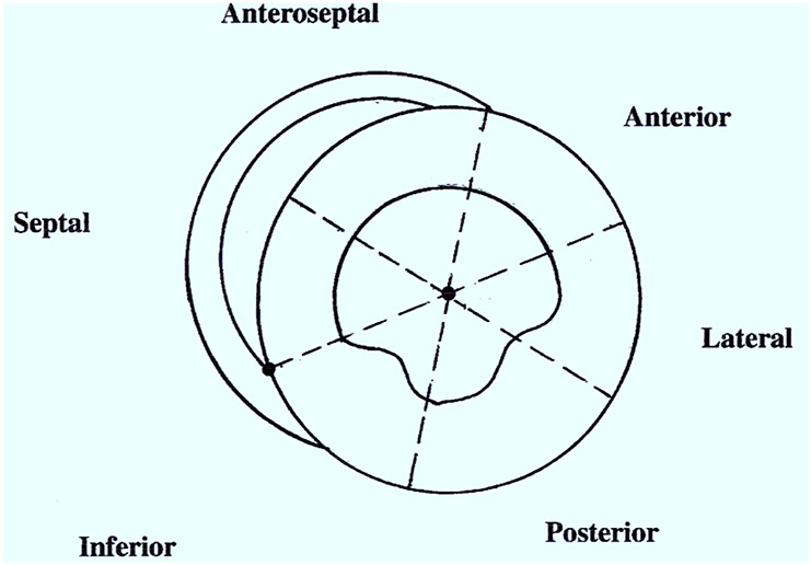
Normal values for regional left ventricular wall motion, although documented in adults, have not been reported in healthy newborns.

METHODS

This study prospectively evaluated global and segmental systolic and diastolic cardiac function by color kinesis in clinically asymptomatic healthy newborns.

RESULTS

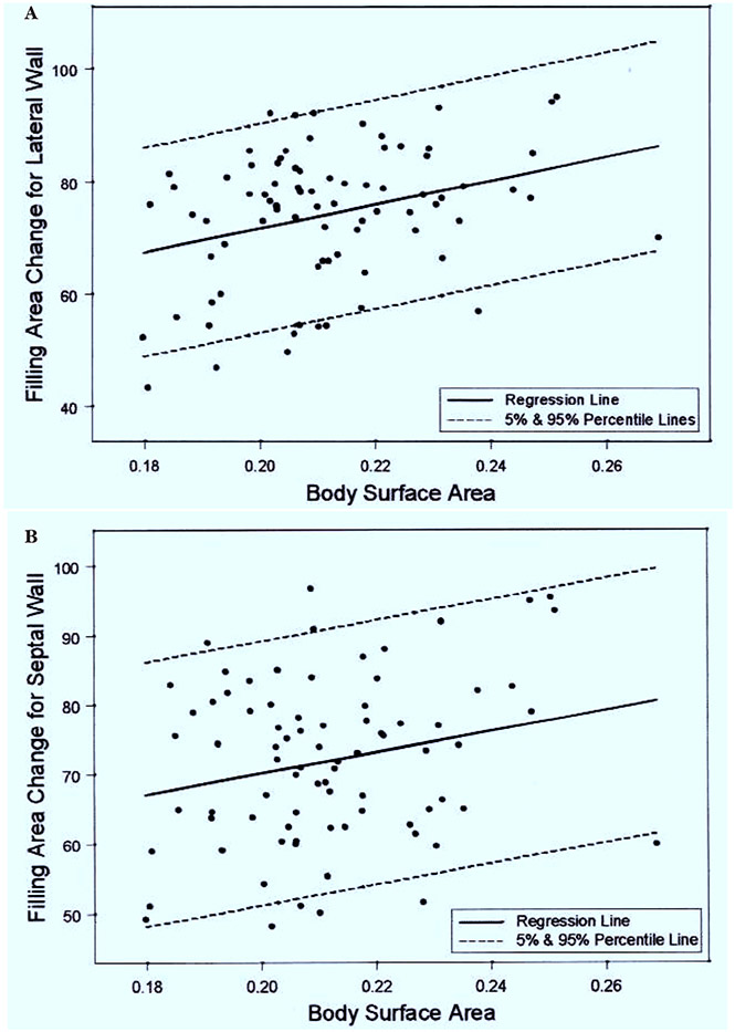
Eighty-eight asymptomatic infants who were less than 48 hours old were studied. Systolic and diastolic parameters of global and regional left ventricular function are reported as means +/- SD, medians, 5th and 95th percentiles to establish the normative values for newborns. The reported fractional area changes during systole and diastole are similar to the reported normal values for older subjects. Higher body surface area significantly correlated with an increased peak velocity during systole, and fractional area changes during filling of the lateral wall.

CONCLUSIONS

Our report of left ventricular regional wall-motion characteristics of healthy newborns, as evaluated by color kinesis, may help in the objective evaluation and management of newborns suspected to have global or segmental ventricular dysfunction.

摘要

背景

尽管已记录了成人左心室壁节段运动的正常数值,但健康新生儿的相关数值尚未见报道。

方法

本研究采用彩色室壁运动技术对临床无症状的健康新生儿进行前瞻性评估,分析全心和节段性的心脏收缩及舒张功能。

结果

对88例年龄小于48小时的无症状婴儿进行了研究。以平均值±标准差、中位数、第5和第95百分位数报告全心和左心室节段功能的收缩期和舒张期参数,以确立新生儿的正常数值。所报告的收缩期和舒张期面积变化分数与年龄较大受试者的报告正常值相似。较高的体表面积与收缩期峰值速度增加以及侧壁充盈期面积变化分数增加显著相关。

结论

我们通过彩色室壁运动技术评估健康新生儿左心室节段壁运动特征的报告,可能有助于对疑似存在全心或节段性心室功能障碍的新生儿进行客观评估和管理。

https://cdn.ncbi.nlm.nih.gov/pmc/blobs/5346/10273883/964a778b7742/nihms-1901990-f0002.jpg
https://cdn.ncbi.nlm.nih.gov/pmc/blobs/5346/10273883/166dc8578447/nihms-1901990-f0001.jpg
https://cdn.ncbi.nlm.nih.gov/pmc/blobs/5346/10273883/964a778b7742/nihms-1901990-f0002.jpg
https://cdn.ncbi.nlm.nih.gov/pmc/blobs/5346/10273883/166dc8578447/nihms-1901990-f0001.jpg
https://cdn.ncbi.nlm.nih.gov/pmc/blobs/5346/10273883/964a778b7742/nihms-1901990-f0002.jpg

相似文献

1
Quantification of regional left ventricular wall motion in newborns by color kinesis.应用彩色室壁运动技术定量分析新生儿左心室壁节段运动
J Am Soc Echocardiogr. 2002 Apr;15(4):356-63. doi: 10.1067/mje.2002.117295.
2
Quantitative evaluation of global and regional left ventricular diastolic function with color kinesis.应用彩色室壁运动技术对左心室整体及局部舒张功能进行定量评估。
Circulation. 1998 Mar 24;97(11):1053-61. doi: 10.1161/01.cir.97.11.1053.
3
Diastolic filling abnormalities by color kinesis in newborns exposed to intrauterine cocaine.应用彩色室壁运动分析宫内暴露于可卡因的新生儿的舒张期充盈异常。
J Am Soc Echocardiogr. 2002 May;15(5):447-53. doi: 10.1067/mje.2002.117296.
4
Evaluation of regional left ventricular wall motion with color kinesis: comparison with two-dimensional echocardiography in patients after acute myocardial infarction.应用彩色室壁运动技术评估局部左心室壁运动:与急性心肌梗死后患者二维超声心动图的比较
G Ital Cardiol. 1998 Sep;28(9):984-95.
5
Segmental analysis of color kinesis images: new method for quantification of the magnitude and timing of endocardial motion during left ventricular systole and diastole.彩色室壁运动分析图像的节段分析:定量评估左心室收缩期和舒张期内心内膜运动幅度和时间的新方法。
Circulation. 1997 Apr 15;95(8):2082-97. doi: 10.1161/01.cir.95.8.2082.
6
Effects of inotropic stimulation on segmental left ventricular relaxation quantified by color kinesis.通过彩色室壁运动分析评估变力性刺激对左心室节段舒张功能的影响。
Am J Cardiol. 2000 Jun 15;85(12):1476-80. doi: 10.1016/s0002-9149(00)00798-0.
7
Echocardiographic quantification of regional left ventricular wall motion with color kinesis.应用彩色室壁运动技术对左心室壁节段运动进行超声心动图定量分析。
Circulation. 1996 May 15;93(10):1877-85. doi: 10.1161/01.cir.93.10.1877.
8
Quantification of the myocardial velocity gradient and myocardial wall thickening velocity in healthy children: a new indicator of regional myocardial wall motion.健康儿童心肌速度梯度和心肌壁增厚速度的量化:区域心肌壁运动的新指标。
J Am Soc Echocardiogr. 2002 Jun;15(6):624-32. doi: 10.1067/mje.2002.118241.
9
Normal values of regional left ventricular endocardial motion: multicenter color kinesis study.局部左心室心内膜运动的正常值:多中心彩色室壁运动分析研究
Am J Physiol Heart Circ Physiol. 2000 Nov;279(5):H2464-76. doi: 10.1152/ajpheart.2000.279.5.H2464.
10
Assessment of left ventricular regional wall motion by color kinesis technique: comparison with angiographic findings.应用彩色室壁运动技术评估左心室局部室壁运动:与血管造影结果的比较。
Echocardiography. 2000 Aug;17(6 Pt 1):521-7. doi: 10.1046/j.1540-8175.2000.00521.x.

引用本文的文献

1
Automatic classification of left ventricular regional wall motion abnormalities in echocardiography images using nonrigid image registration.利用非刚性图像配准技术对超声心动图图像中的左心室局部壁运动异常进行自动分类。
J Digit Imaging. 2013 Oct;26(5):909-19. doi: 10.1007/s10278-012-9543-x.
2
Left ventricle wall motion quantification from echocardiographic images by non-rigid image registration.利用超声心动图图像的非刚性图像配准进行左心室壁运动定量分析。
Int J Comput Assist Radiol Surg. 2012 Sep;7(5):769-83. doi: 10.1007/s11548-012-0786-2. Epub 2012 Jul 31.

本文引用的文献

1
Diastolic filling abnormalities by color kinesis in newborns exposed to intrauterine cocaine.应用彩色室壁运动分析宫内暴露于可卡因的新生儿的舒张期充盈异常。
J Am Soc Echocardiogr. 2002 May;15(5):447-53. doi: 10.1067/mje.2002.117296.
2
Normal values of regional left ventricular endocardial motion: multicenter color kinesis study.局部左心室心内膜运动的正常值:多中心彩色室壁运动分析研究
Am J Physiol Heart Circ Physiol. 2000 Nov;279(5):H2464-76. doi: 10.1152/ajpheart.2000.279.5.H2464.
3
Evaluation of regional wall motion and quantitative measures of ventricular function during dobutamine stress echocardiography in pediatric cardiac transplantation patients.
小儿心脏移植患者多巴酚丁胺负荷超声心动图检查时局部室壁运动及心室功能定量指标的评估。
J Am Soc Echocardiogr. 2000 Oct;13(10):932-40. doi: 10.1067/mje.2000.106905.
4
Diastolic biomechanics in normal infants utilizing MRI tissue tagging.利用MRI组织标记技术研究正常婴儿的舒张期生物力学。
Circulation. 2000 Jul 11;102(2):218-24. doi: 10.1161/01.cir.102.2.218.
5
Evaluation of myocardial perfusion using positron emission tomography in infants following a neonatal arterial switch operation.新生儿动脉调转手术后婴儿心肌灌注的正电子发射断层扫描评估
Pediatr Cardiol. 2000 Mar-Apr;21(2):111-8. doi: 10.1007/s002469910015.
6
Regional differences in shape and load in normal and diseased hearts studied by three dimensional tagged magnetic resonance imaging.通过三维标记磁共振成像研究正常和患病心脏的形状及负荷的区域差异。
Int J Card Imaging. 1999 Aug;15(4):309-21. doi: 10.1023/a:1006132709895.
7
Objective evaluation of regional left ventricular wall motion during dobutamine stress echocardiographic studies using segmental analysis of color kinesis images.使用彩色室壁运动分析图像的节段分析,在多巴酚丁胺负荷超声心动图研究期间对局部左心室壁运动进行客观评估。
J Am Coll Cardiol. 1999 Aug;34(2):409-19. doi: 10.1016/s0735-1097(99)00233-8.
8
Abnormal left ventricular filling after neonatal repair of congenital heart disease: association with increased mortality and morbidity.先天性心脏病新生儿修复术后左心室充盈异常:与死亡率和发病率增加的关联。
Am Heart J. 1998 Dec;136(6):1075-80. doi: 10.1016/s0002-8703(98)70166-8.
9
Use of color kinesis for evaluation of left ventricular filling in patients with dilated cardiomyopathy and mitral regurgitation.应用彩色室壁运动分析技术评估扩张型心肌病合并二尖瓣反流患者的左心室充盈情况。
J Am Coll Cardiol. 1998 Jun;31(7):1598-606. doi: 10.1016/s0735-1097(98)00144-2.
10
Quantitative evaluation of global and regional left ventricular diastolic function with color kinesis.应用彩色室壁运动技术对左心室整体及局部舒张功能进行定量评估。
Circulation. 1998 Mar 24;97(11):1053-61. doi: 10.1161/01.cir.97.11.1053.