Poulter L W, Lehner T
Department of Immunology, Royal Free Hospital and School of Medicine, London, England.
Clin Exp Immunol. 1989 Nov;78(2):189-95.
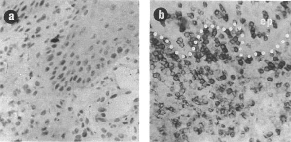
A qualitative and quantitative immunohistological investigation was performed on biopsies of oral ulcers from patients with Behcet's syndrome (BS) and those with recurrent oral ulcers (ROU). The results were compared with control oral biopsies from patients with other diseases and normal oral mucosa. The expression of HLA-DR on the cell membrane of keratinocytes was found in 13 out of 15 lesions from patients with BS and ROU, as compared with only one out of 15 controls. The relative density of HLA-DR was investigated quantitatively by microdensitometry and this confirmed that DR expression in the epithelial cells of patients with BS and ROU was significantly greater than in diseased and normal control oral tissues. A prominent mononuclear cell infiltration consisted predominantly of T lymphocytes and mature macrophages. Analysis of the CD4 and CD8 subsets of T cells failed to show significant differences between BS, ROU and control diseased tissues. Increased numbers of Langerhans cells were found in the epithelium by morphometric analysis with the CD1 monoclonal antibody in BS and ROU but an increased number was also found in lichen planus. The results suggest that the immunohistological changes in oral lesions of BS and ROU manifest an enhanced immune response in the epithelium, keratinocytes express HLA-class II antigen and increased number of Langerhans cells as well as in the lamina propria with a prominent infiltration of CD4, CD8 and macrophage-like cells. The characteristic pattern of exacerbations and remissions of oral ulceration can be interpreted by the hypothesis that an initiating microbial agent may induce a mononuclear cell infiltration, with the release of cytokines, expression of class II antigen in keratinocytes and causing ulceration, followed by down-regulation of immunity by tolerant T cells induced by the class II positive keratinocytes, leading to a remission.
对贝赫切特综合征(BS)患者和复发性口腔溃疡(ROU)患者的口腔溃疡活检标本进行了定性和定量免疫组织学研究。将结果与其他疾病患者的对照口腔活检标本和正常口腔黏膜进行比较。在15例BS和ROU患者的病变中,有13例发现角质形成细胞膜上有HLA-DR表达,而15例对照中只有1例有该表达。通过显微密度测定法定量研究了HLA-DR的相对密度,这证实了BS和ROU患者上皮细胞中的DR表达明显高于患病和正常对照口腔组织。显著的单核细胞浸润主要由T淋巴细胞和成熟巨噬细胞组成。对T细胞的CD4和CD8亚群分析未能显示BS、ROU和对照患病组织之间有显著差异。通过用CD1单克隆抗体进行形态计量分析,发现BS和ROU患者上皮中朗格汉斯细胞数量增加,但扁平苔藓中也发现数量增加。结果表明,BS和ROU口腔病变中的免疫组织学变化表现为上皮中免疫反应增强,角质形成细胞表达HLA-II类抗原,朗格汉斯细胞数量增加,固有层中CD4、CD8和巨噬细胞样细胞浸润显著。口腔溃疡发作和缓解的特征模式可以用以下假说来解释:一种起始微生物因子可能诱导单核细胞浸润,释放细胞因子,角质形成细胞中II类抗原表达并导致溃疡形成,随后由II类阳性角质形成细胞诱导的耐受T细胞使免疫下调,导致缓解。